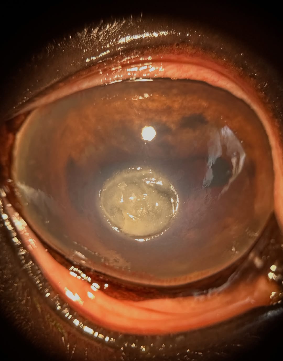
QuikVue Vet Case Share - fungal keratitis
We are glad to share a vet case study captured by QuikVue eye imaging adaptor from Dr. Allison Fuchs.
Another horse with fungal keratitis! I had 4 cases in a week and man, this is a frustrating disease for a veterinary ophthalmologist.
Horses are prone to corneal ulcers from trauma due to their environment and big eyes, and fungal infection setting in is common in some parts of the world - many of them! In our area of the east coast it is incredibly prevalent and I am a firm believer that ANY horse with a corneal ulcer should have topical antifungal as part of the treatment.
This one has me pretty concerned. You can see the start of a moat or furrow at the top right of the lesion, a deep area that is generally associated with rapid corneal damage occurring. This gives away how deep the lesion really is even though the rest of it doesn’t look too bad - most of that is necrotic cornea and inflammatory debris.
We are treating this horse aggressively with a subpalpebral lavage system, frequent topical antibiotics, antifungals, and anti collagenases, and medication to help with discomfort and inflammation. Hopefully we can turn this around, but fungal keratitis in horses can have a guarded prognosis.
 | 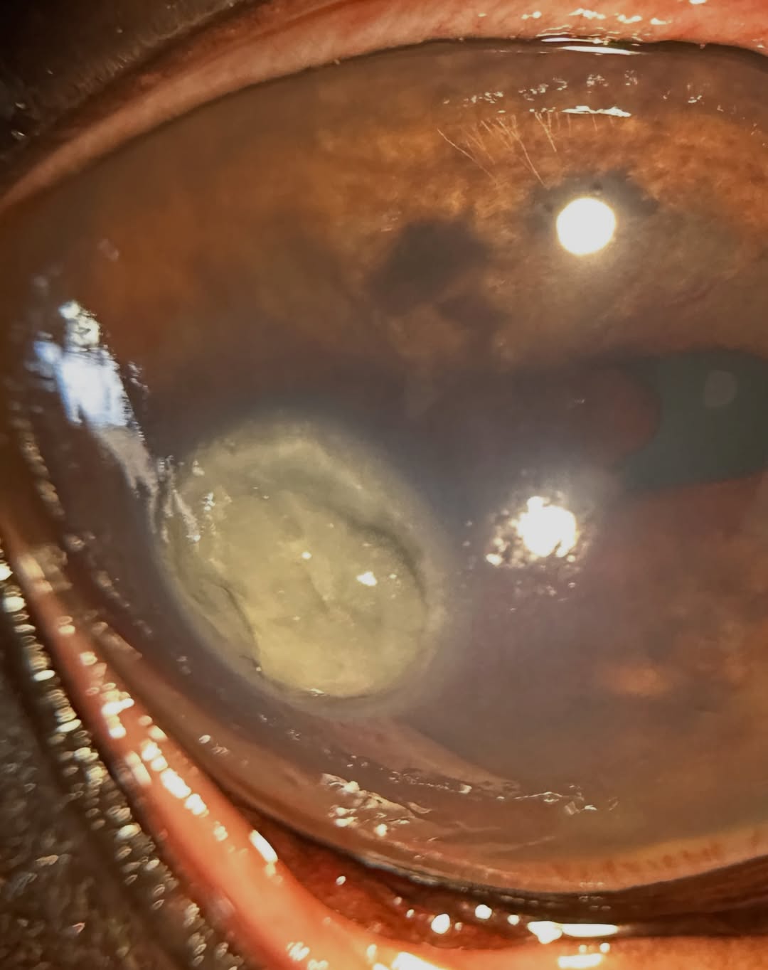 |
We are glad to share a vet case study captured by QuikVue eye imaging adaptor from Dr. Allison Fuchs.
Another horse with fungal keratitis! I had 4 cases in a week and man, this is a frustrating disease for a veterinary ophthalmologist.
Horses are prone to corneal ulcers from trauma due to their environment and big eyes, and fungal infection setting in is common in some parts of the world - many of them! In our area of the east coast it is incredibly prevalent and I am a firm believer that ANY horse with a corneal ulcer should have topical antifungal as part of the treatment.
This one has me pretty concerned. You can see the start of a moat or furrow at the top right of the lesion, a deep area that is generally associated with rapid corneal damage occurring. This gives away how deep the lesion really is even though the rest of it doesn’t look too bad - most of that is necrotic cornea and inflammatory debris.
We are treating this horse aggressively with a subpalpebral lavage system, frequent topical antibiotics, antifungals, and anti collagenases, and medication to help with discomfort and inflammation. Hopefully we can turn this around, but fungal keratitis in horses can have a guarded prognosis.
| |