Upgrade Your Slit Lamp to Digital Imaging System
Slit-lamp examination is still where much of anterior segment care happens. On a traditional slit lamp, the clinician sees findings through the eyepiece—but the instrument itself does not capture photo or video. What you observe during the exam often stays in the moment: there is no image saved to the chart, no clip for follow-up comparison, and no visual record you can show the patient on screen.
That limit shows up in daily workflow. Follow-up visits may depend on notes, memory, or verbal description rather than the same view you had at the lamp. Patient education can be harder when you cannot point to a stored image of staining, tear film, or anterior segment findings. Staff may re-explain findings without a shared visual reference from the prior visit.
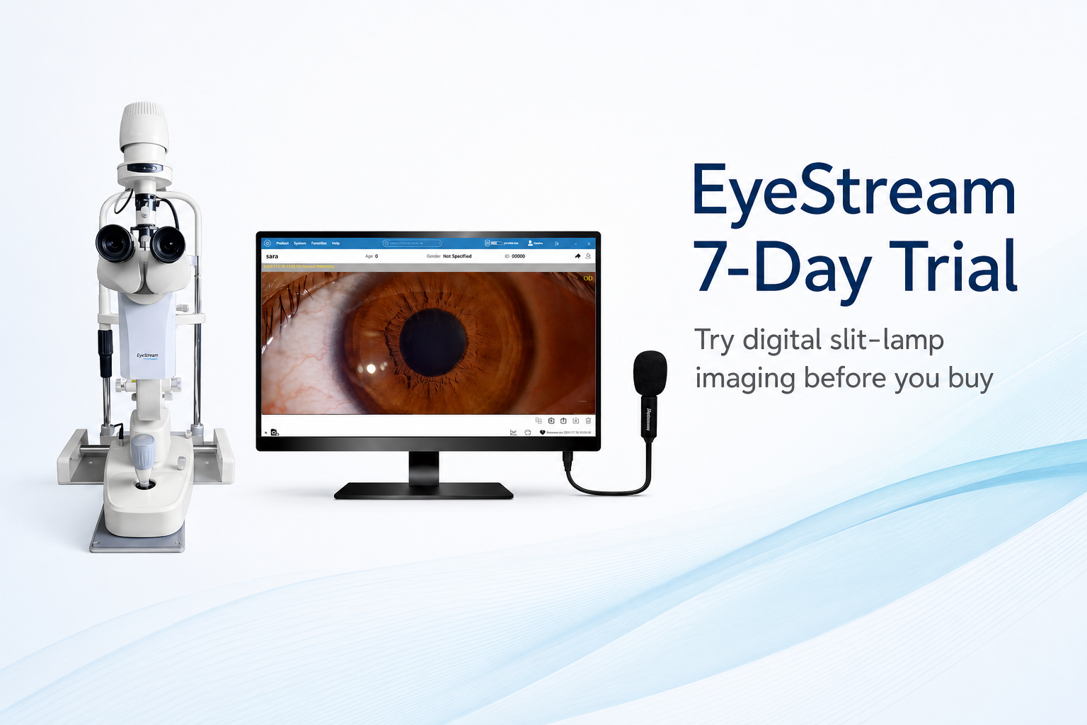
Many practices want image-based documentation at the slit lamp—not a separate imaging step after the exam, and not a full system replacement. EyeStream is a compact camera module from VisuScience designed to add digital photo and video to compatible slit lamps, with capture and review in VisuDoc software—without replacing the full slit-lamp system.
Built for the Slit Lamp Already in Your Room
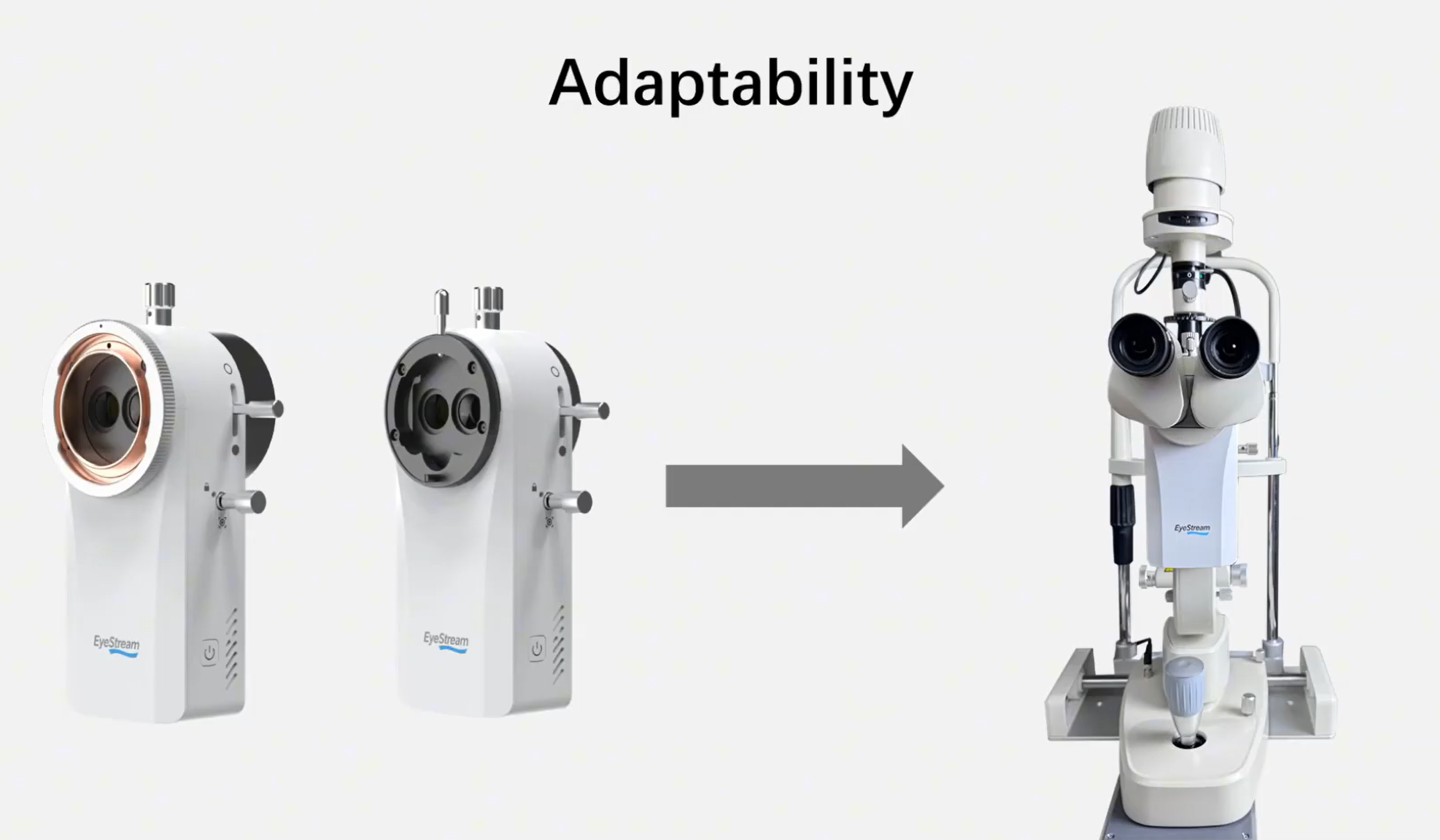
Most traditional slit lamps in daily use were built for eyepiece examination—not built-in photo or video. Replacing every lamp in the room to gain imaging is often not practical. Capital planning often favors upgrades that extend existing equipment rather than replace entire platforms.
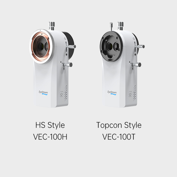
EyeStream is offered in two dedicated models—one designed for Haag Streit slit lamps and one for Topcon-type slit lamps. Each configuration is built to fit upper light source slit lamps at 3× and 5× magnification, so practices can match the module to the lamp already in the room.
Before you plan installation, confirm lamp model, magnification configuration, mount style, and regional authorization for your site.
VisuDoc Workflow: Photo, Video, and Hands-Free Control
Traditional slit lamps leave documentation to notes and recall. If adding capture means leaving the eyepiece or breaking examination flow, adoption often stalls.
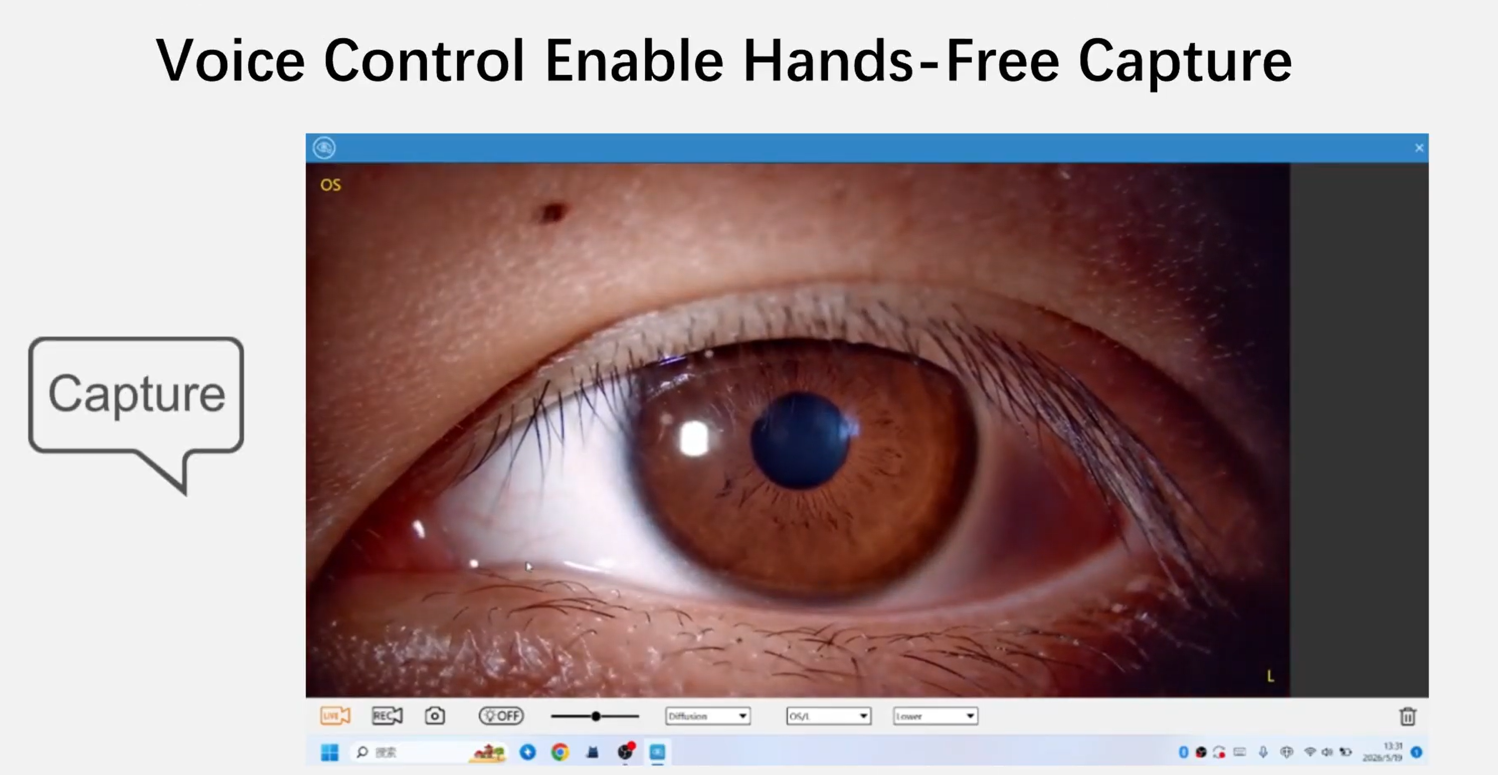
Photo and video capture take place in VisuDoc software. With the VisuScience speaker connected to the workstation, clinicians can use voice control during slit-lamp examination—for example, say “capture” for a still saved to the active patient record, “video” to record, and “stop” to end. Say “meibo,” “slit,” or “diffusion” to change imaging modes on screen. Command words must match shipped VisuDoc UI, IFU, and regional authorization.
Captures can save automatically to the patient record you have open in VisuDoc—turning eyepiece-only observation into stored images and video for follow-up comparison and patient discussion. EyeStream is designed to support workflow at the point of care; it does not replace clinical judgment.
- Voice-controlled still capture and video recording in VisuDoc
- Mode switching during exam (meibo, slit, diffusion—per authorized software)
- Stills and clips saved to the open patient record during the visit
Fluorescein Imaging, Patient Education, and Next Steps
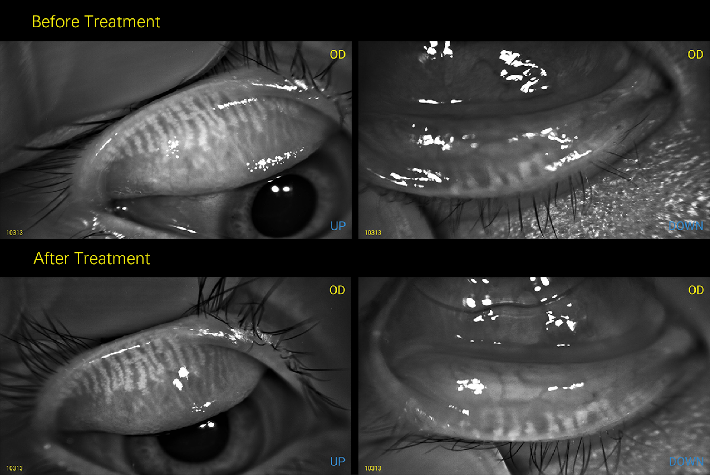
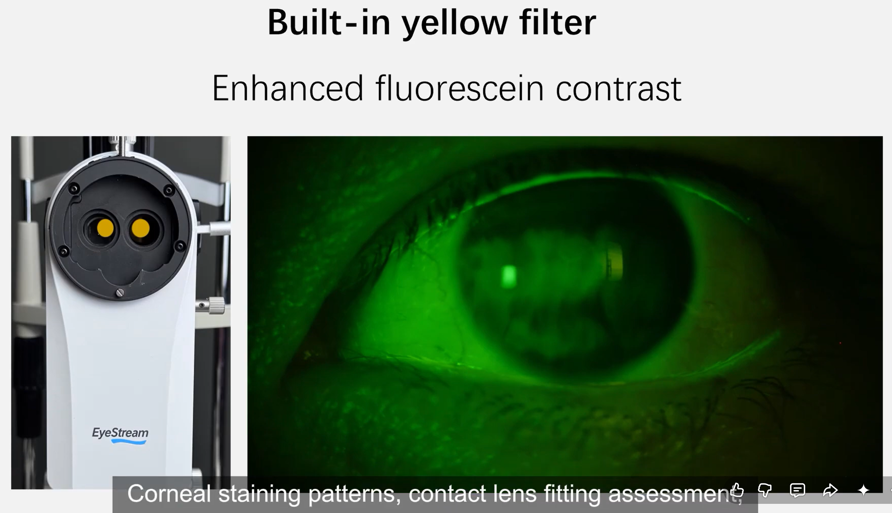
Fluorescein staining, contact lens fitting assessment, and tear film breakup observation are routine slit-lamp tasks—but on a traditional lamp, enhanced views at the eyepiece do not automatically become images in the patient record.
EyeStream includes a built-in yellow filter designed to enhance fluorescein contrast during slit-lamp examination. Corneal staining patterns, contact lens fitting assessment, and tear film breakup observation can be captured and stored in VisuDoc—without adding a separate filter to your workflow. Images and clips may support patient education and longitudinal review when used according to your care plan and local practice standards.
EyeStream is intended to support image-based documentation, patient discussion, and follow-up imaging at the point of care. The practical takeaway: keep the slit lamp you already use, add photo and video at the exam, and confirm fit and authorization for your configuration before adoption.
Learn more at www.visuscience.com
Slit-lamp examination is still where much of anterior segment care happens. On a traditional slit lamp, the clinician sees findings through the eyepiece—but the instrument itself does not capture photo or video. What you observe during the exam often stays in the moment: there is no image saved to the chart, no clip for follow-up comparison, and no visual record you can show the patient on screen.
That limit shows up in daily workflow. Follow-up visits may depend on notes, memory, or verbal description rather than the same view you had at the lamp. Patient education can be harder when you cannot point to a stored image of staining, tear film, or anterior segment findings. Staff may re-explain findings without a shared visual reference from the prior visit.
Many practices want image-based documentation at the slit lamp—not a separate imaging step after the exam, and not a full system replacement. EyeStream is a compact camera module from VisuScience designed to add digital photo and video to compatible slit lamps, with capture and review in VisuDoc software—without replacing the full slit-lamp system.
Built for the Slit Lamp Already in Your Room
Most traditional slit lamps in daily use were built for eyepiece examination—not built-in photo or video. Replacing every lamp in the room to gain imaging is often not practical. Capital planning often favors upgrades that extend existing equipment rather than replace entire platforms.
EyeStream is offered in two dedicated models—one designed for Haag Streit slit lamps and one for Topcon-type slit lamps. Each configuration is built to fit upper light source slit lamps at 3× and 5× magnification, so practices can match the module to the lamp already in the room.
Before you plan installation, confirm lamp model, magnification configuration, mount style, and regional authorization for your site.
VisuDoc Workflow: Photo, Video, and Hands-Free Control
Traditional slit lamps leave documentation to notes and recall. If adding capture means leaving the eyepiece or breaking examination flow, adoption often stalls.
Photo and video capture take place in VisuDoc software. With the VisuScience speaker connected to the workstation, clinicians can use voice control during slit-lamp examination—for example, say “capture” for a still saved to the active patient record, “video” to record, and “stop” to end. Say “meibo,” “slit,” or “diffusion” to change imaging modes on screen. Command words must match shipped VisuDoc UI, IFU, and regional authorization.
Captures can save automatically to the patient record you have open in VisuDoc—turning eyepiece-only observation into stored images and video for follow-up comparison and patient discussion. EyeStream is designed to support workflow at the point of care; it does not replace clinical judgment.
-
Voice-controlled still capture and video recording in VisuDoc
-
Mode switching during exam (meibo, slit, diffusion—per authorized software)
-
Stills and clips saved to the open patient record during the visit
Fluorescein Imaging, Patient Education, and Next Steps
Fluorescein staining, contact lens fitting assessment, and tear film breakup observation are routine slit-lamp tasks—but on a traditional lamp, enhanced views at the eyepiece do not automatically become images in the patient record.
EyeStream includes a built-in yellow filter designed to enhance fluorescein contrast during slit-lamp examination. Corneal staining patterns, contact lens fitting assessment, and tear film breakup observation can be captured and stored in VisuDoc—without adding a separate filter to your workflow. Images and clips may support patient education and longitudinal review when used according to your care plan and local practice standards.
EyeStream is intended to support image-based documentation, patient discussion, and follow-up imaging at the point of care. The practical takeaway: keep the slit lamp you already use, add photo and video at the exam, and confirm fit and authorization for your configuration before adoption.
Learn more at www.visuscience.com