QuikVue Vet Case Share - Deep, infected corneal ulcer in a bulldog
We are glad to share a vet case study captured by QuikVue eye imaging adaptor from Dr. Allison Fuchs.
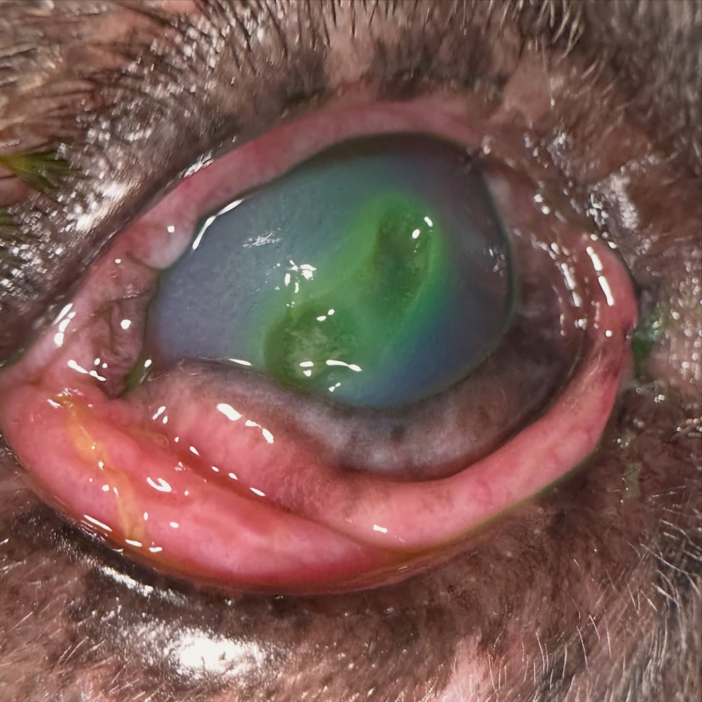
These are always the last appointment of the day before a weekend, and unfortunately this dog really did have the best prognosis with surgery - so to surgery we went! In this case, the patient had been undergoing treatment with a primary care veterinarian for 3 weeks or so and was referred to ophthalmology for a suspected indolent ulcer and debridement. On exam, the dog had an underlying dry eye (KCS), and as you can see, this ulcer is actually very deep with white blood cell infiltrate. It’s hard to tell in the photo but this was also severely keratomalacic (melting).
Dogs with dry eye are much more prone to corneal ulcers, and to those ulcers not healing appropriately. In many cases we can treat medically with antibiotic eye drops and drops to help the cornea heal normally. We elected for surgery to graft the cornea in this case for several reasons - dry eye, a brachycephalic breed, and heading into a severe winter storm over the weekend. The owners didn’t want to take the risk of corneal rupture and not being able to get treatment if the weather precluded travel!
This dog’s surgery went very well and we’re hopeful that it will look good at the progress exam next week, assuming we aren’t all snowed in!
This case highlights the importance of appropriate diagnostics, prompt referral, and client education in veterinary specialty.
We are glad to share a vet case study captured by QuikVue eye imaging adaptor from Dr. Allison Fuchs.
These are always the last appointment of the day before a weekend, and unfortunately this dog really did have the best prognosis with surgery - so to surgery we went! In this case, the patient had been undergoing treatment with a primary care veterinarian for 3 weeks or so and was referred to ophthalmology for a suspected indolent ulcer and debridement. On exam, the dog had an underlying dry eye (KCS), and as you can see, this ulcer is actually very deep with white blood cell infiltrate. It’s hard to tell in the photo but this was also severely keratomalacic (melting).
Dogs with dry eye are much more prone to corneal ulcers, and to those ulcers not healing appropriately. In many cases we can treat medically with antibiotic eye drops and drops to help the cornea heal normally. We elected for surgery to graft the cornea in this case for several reasons - dry eye, a brachycephalic breed, and heading into a severe winter storm over the weekend. The owners didn’t want to take the risk of corneal rupture and not being able to get treatment if the weather precluded travel!
This dog’s surgery went very well and we’re hopeful that it will look good at the progress exam next week, assuming we aren’t all snowed in!
This case highlights the importance of appropriate diagnostics, prompt referral, and client education in veterinary specialty.