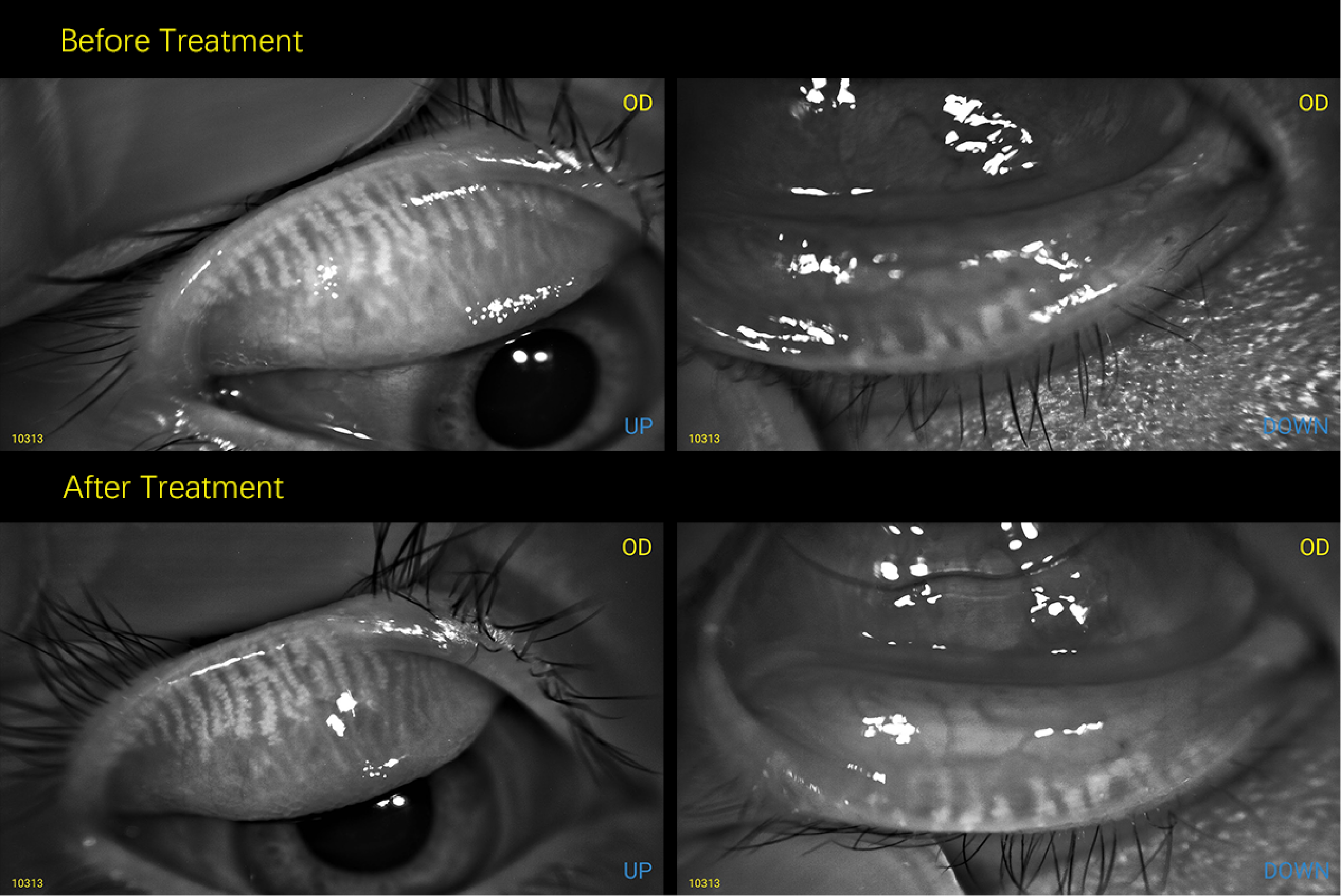
QuikVue Vet Case Share- nasty Enterococcus bacteria
We are glad to share a vet case study captured by QuikVue eye imaging adaptor fromDr. Allison Fuchs.
Case progress updates! This is the dog who grew a nasty Enterococcus bacteria in the corneal sample that I showed recently. These photos cover a span of about 4 weeks, from the initial presentation with a heavily infected stromal abscess which then progressed into a deep ulcer in the second image. That’s when we went to surgery to keratectomy to remove as much infection as possible and grafted in a bioscaffold product to add support. She already had lots of blood vessels, so a conjunctival graft was not what I chose to perform in this instance.
We also cultured tissue from surgery to alter her medication, and one week later the infection was well controlled and the graft had vascularized in the third image. Now 3 weeks out from surgery the graft is scarring down and I can’t wait to show you another progress update in another few weeks - you’ll be amazed how good she’s going to look!
 |  |
We are glad to share a vet case study captured by QuikVue eye imaging adaptor fromDr. Allison Fuchs.
Case progress updates! This is the dog who grew a nasty Enterococcus bacteria in the corneal sample that I showed recently. These photos cover a span of about 4 weeks, from the initial presentation with a heavily infected stromal abscess which then progressed into a deep ulcer in the second image. That’s when we went to surgery to keratectomy to remove as much infection as possible and grafted in a bioscaffold product to add support. She already had lots of blood vessels, so a conjunctival graft was not what I chose to perform in this instance.
We also cultured tissue from surgery to alter her medication, and one week later the infection was well controlled and the graft had vascularized in the third image. Now 3 weeks out from surgery the graft is scarring down and I can’t wait to show you another progress update in another few weeks - you’ll be amazed how good she’s going to look!
| |