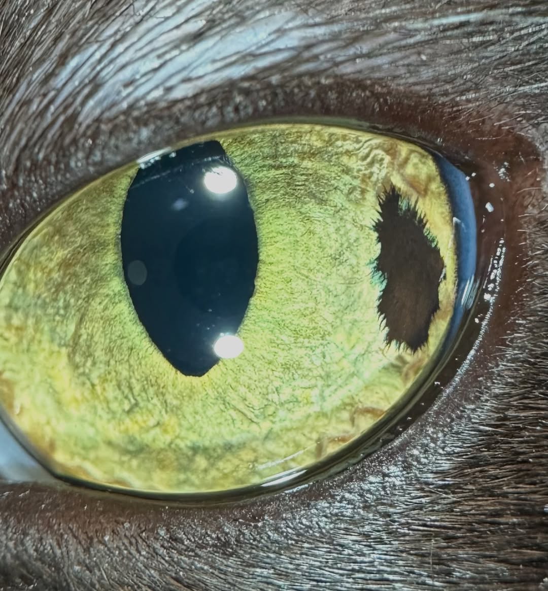
QuikVue Vet Case Share - progressive iris pigmentation
We are glad to share a vet case study captured by QuikVue eye imaging adaptor from Dr. Allison Fuchs.
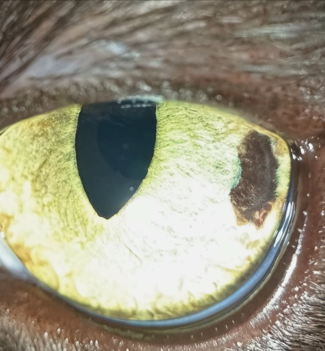
One of my favorite conditions to be able to offer treatment for is feline diffuse iris melanosis, when we catch things early like this cat.
This is a young cat with progressive iris pigmentation. The intraocular pressure is normal and there is no active inflammation inside the eye. He is visual and comfortable. What is this and what do we do about it? This condition can be challenging because we have so little solid information about how it behaves! This is FDIM, or feline diffuse iris melanosis/melanoma. There is no real way to tell without histopathology whether the change is benign or malignant, but we evaluate for certain characteristics that make us more or less concerned. A young cat with velvety pigmentation that is progressively expanding makes me worried. There are reports of metastatic disease from iris melanoma in cats, but the reported rates range from 16-63% which is wildly unhelpful. More recent studies show lower rates. Some people advocate for early enucleation. Personally I offer a range of options depending on the cat's age, rate of progression, the appearance of the lesions, and the owner's motivation. We can monitor (watch and wait), take an iris biopsy, perform trans-corneal laser ablation to try to slow down the growth, or remove the eye. I also offer staging (chest x-rays and abdominal ultrasound) prior to surgery, especially in more advanced cases.
This case underwent staging and laser, and the second image is about 2 weeks post laser. If you look carefully, you'll notice that it's less dark and more grey, and the edges have contracted down. The goal is to kill the melanocytes and prevent them from growing! Hopefully this area will continue to regress, but we monitor these cats closely.
 |  |
We are glad to share a vet case study captured by QuikVue eye imaging adaptor from Dr. Allison Fuchs.
One of my favorite conditions to be able to offer treatment for is feline diffuse iris melanosis, when we catch things early like this cat.
This is a young cat with progressive iris pigmentation. The intraocular pressure is normal and there is no active inflammation inside the eye. He is visual and comfortable. What is this and what do we do about it? This condition can be challenging because we have so little solid information about how it behaves! This is FDIM, or feline diffuse iris melanosis/melanoma. There is no real way to tell without histopathology whether the change is benign or malignant, but we evaluate for certain characteristics that make us more or less concerned. A young cat with velvety pigmentation that is progressively expanding makes me worried. There are reports of metastatic disease from iris melanoma in cats, but the reported rates range from 16-63% which is wildly unhelpful. More recent studies show lower rates. Some people advocate for early enucleation. Personally I offer a range of options depending on the cat's age, rate of progression, the appearance of the lesions, and the owner's motivation. We can monitor (watch and wait), take an iris biopsy, perform trans-corneal laser ablation to try to slow down the growth, or remove the eye. I also offer staging (chest x-rays and abdominal ultrasound) prior to surgery, especially in more advanced cases.
This case underwent staging and laser, and the second image is about 2 weeks post laser. If you look carefully, you'll notice that it's less dark and more grey, and the edges have contracted down. The goal is to kill the melanocytes and prevent them from growing! Hopefully this area will continue to regress, but we monitor these cats closely.
| |