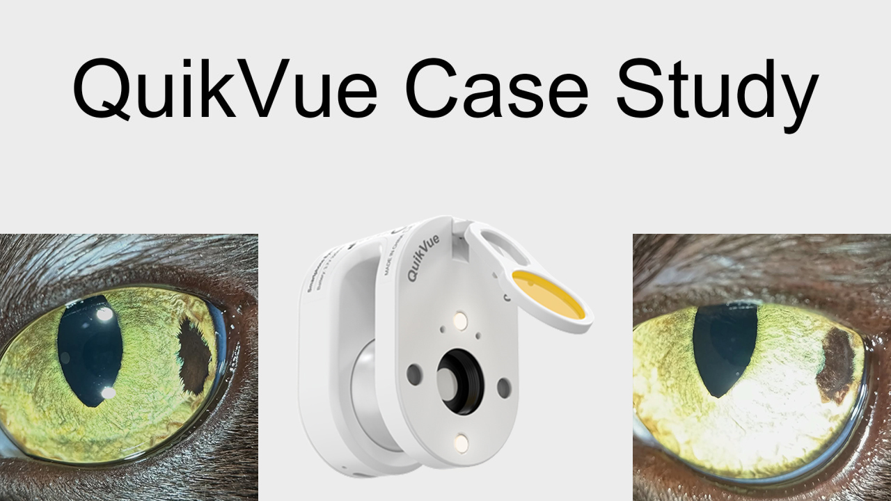
QuikVue Vet Case Share - lens luxation case
We are glad to share a vet case study captured by QuikVue eye imaging adaptor from Dr. Allison Fuchs.
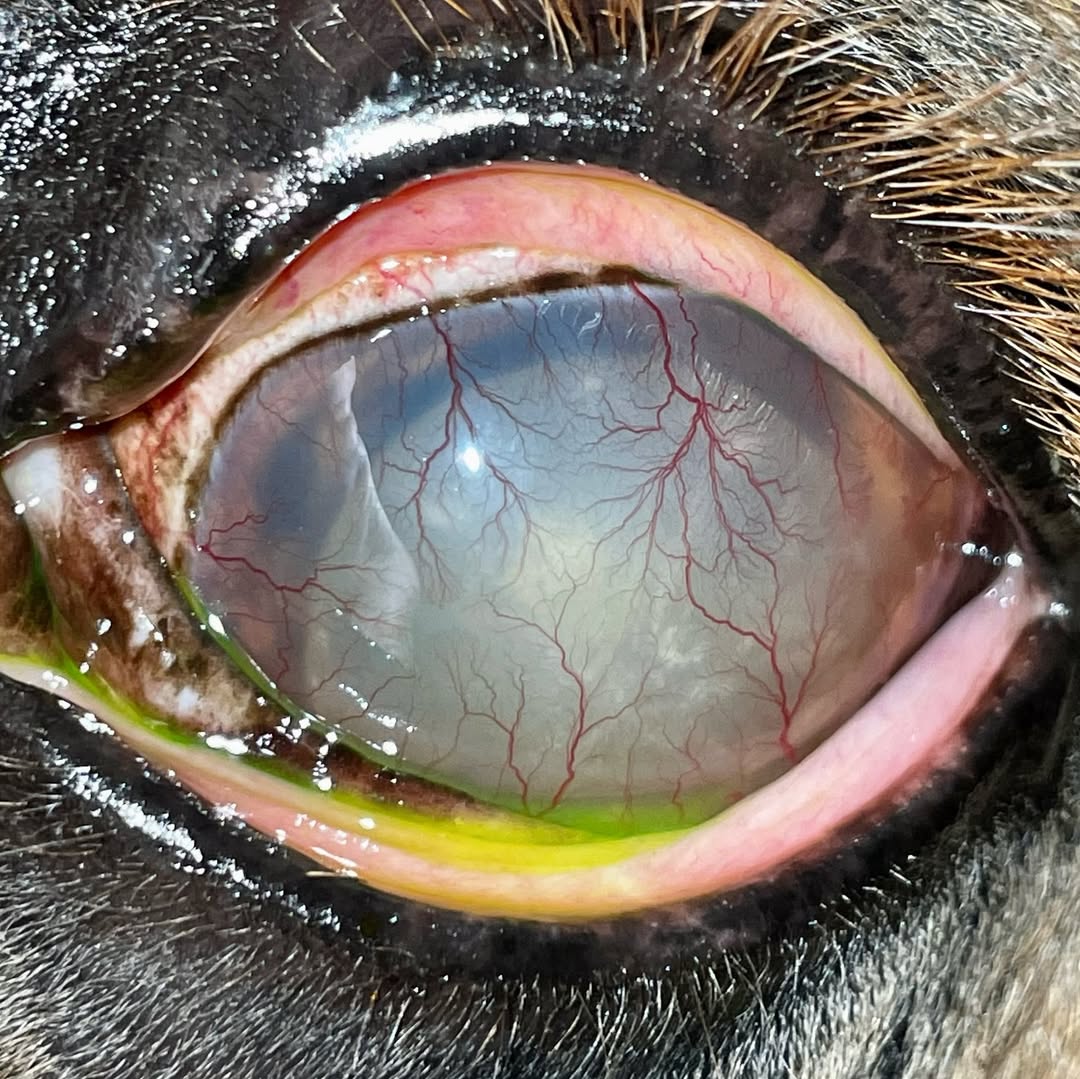
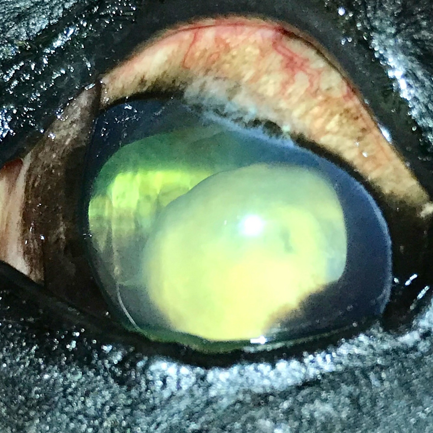
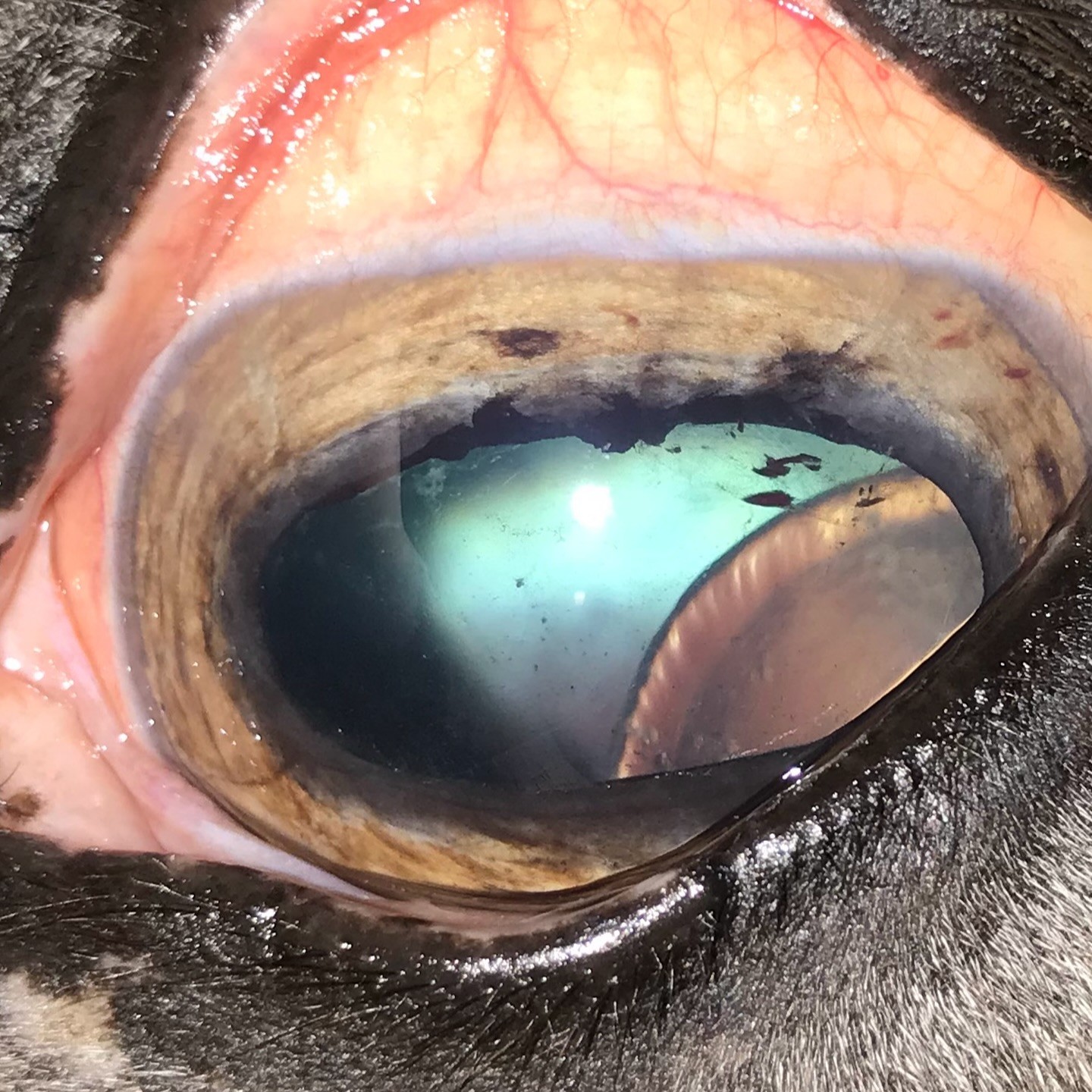
Time to talk about equine lenses! Swipe to check out three very different lens luxation cases in 3 horses.
The crystalline lens is the structure in the middle of the pupil that focuses light on the retina; this is where cataracts form! The lens is held in place by little fibers called zonules that go 360 around the perimeter of the lens like the springs on a trampoline. Dogs can have a primary lens luxation where the fibers break down early.
Horses really only get lens luxations due to chronic uveitis (inflammation) in the eye, or sometimes due to trauma. Trauma generally causes major other issues - but not always! All of these factors make horses poor candidates for surgical lens removal like we perform in dogs. Data is extremely limited, but the prognosis is poor. We generally don’t even have the opportunity to consider a vision-sparing surgery.
Two of these horses have lens luxations due to chronic uveitis, while one had a blunt trauma to the eye that luxated his lens. Surprisingly, the blunt traumatic luxation horse maintained some vision for a long time - he belonged to a personal friend so I got to follow his progress!
 |  |  |