QuikVue Vet Case Share-corneal ulceration
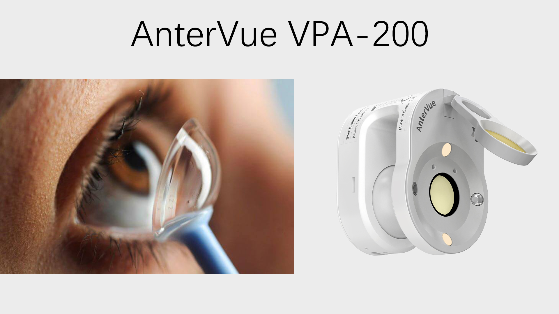
We are glad to share a vet case study captured by QuikVue eye imaging adaptor from Dr.Allison Fuchs.
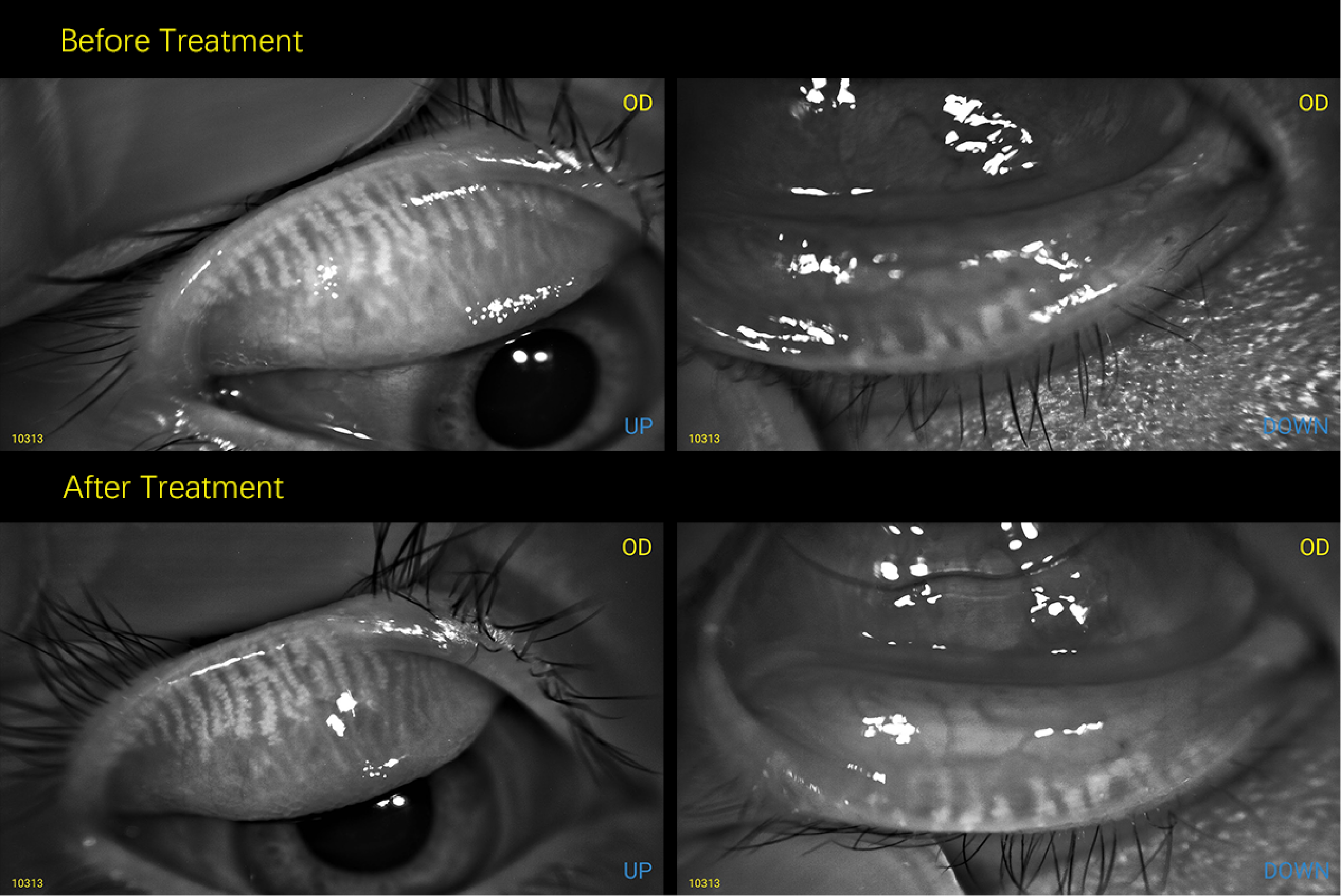
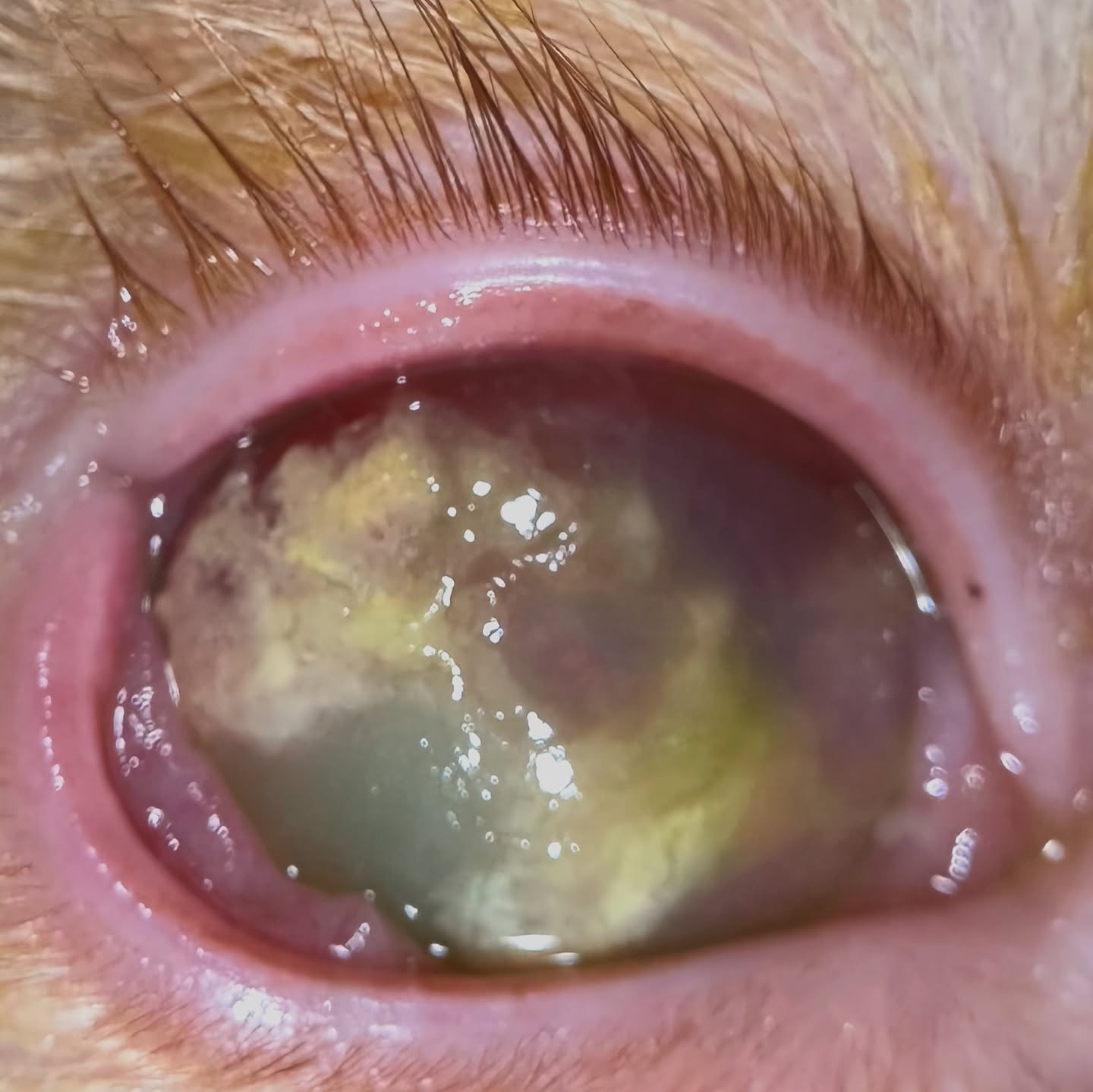
This sweet boy was referred for worsening issues with corneal ulceration which have progressed from one eye to both eyes. Fortunately, these lesions have an explanation and are generally very treatable!
This cat was diagnosed with a classic case of Eosinophilic Keratitis (EK), which is a chronic inflammatory disease of the ocular surface. This primarily happens in cats, though we can also see it in horses and some other small animals.
EK is an immune mediated disease that causes eosinophils, a white blood cell, to invade the cornea. They can have mild corneal vascularization or be as severe as this cat’s extensive plaques. We confirmed the diagnosis with corneal cytology to look for eosinophils and will treat him with topical Prednisolone, cyclosporin, continue some topical antibiotics, and hopefully at his next recheck this will look amazing. Every individual is a little bit different so we may need to change our medication depending on his response!
 |  |
We are glad to share a vet case study captured by QuikVue eye imaging adaptor from Dr.Allison Fuchs.
This sweet boy was referred for worsening issues with corneal ulceration which have progressed from one eye to both eyes. Fortunately, these lesions have an explanation and are generally very treatable!
This cat was diagnosed with a classic case of Eosinophilic Keratitis (EK), which is a chronic inflammatory disease of the ocular surface. This primarily happens in cats, though we can also see it in horses and some other small animals.
EK is an immune mediated disease that causes eosinophils, a white blood cell, to invade the cornea. They can have mild corneal vascularization or be as severe as this cat’s extensive plaques. We confirmed the diagnosis with corneal cytology to look for eosinophils and will treat him with topical Prednisolone, cyclosporin, continue some topical antibiotics, and hopefully at his next recheck this will look amazing. Every individual is a little bit different so we may need to change our medication depending on his response!
| |