QuikVue Vet Case Share-corneal calcific degeneration
We are glad to share a vet case study captured by QuikVue eye imaging adaptor from Dr. Allison Fuchs.
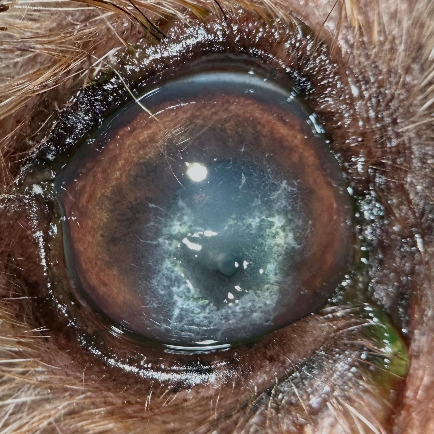
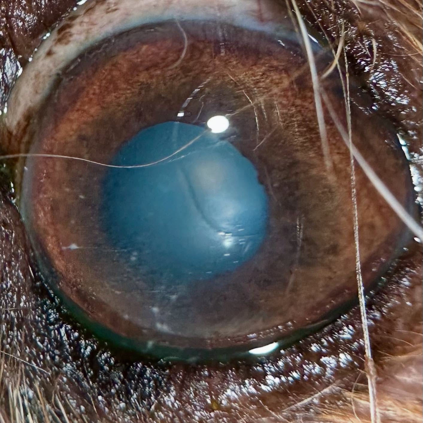
What’s going on here? This is one of the most frustrating conditions for me. This older dog was referred for a deep corneal ulcer, and you can see the white speckling all around the corneal defect in the first image. It’s also there in the second photo, but more subtle. This is corneal calcific degeneration. The white is calcium deposits, which happens due to age related changes in corneal metabolism, prior corneal trauma, underlying systemic metabolic disorders, or sometimes inherited reasons.
Calcium does not belong in the cornea, and the crystals can poke through the epithelium causing pain, squinting, and ocular discharge. At worst, we end up like this dog, and large areas of abnormally calcified cornea flake off into deep ulcers that have a high risk for corneal rupture. These ulcers tend not to heal well because the cornea is abnormal. Surgery is often necessary, but many of these dogs are poor candidates for anesthesia.
For this case, we are going to try to treat medically and hope the ulcer doesn’t rupture. The dog will be on topical antibiotics, as well as a topical EDTA to try to bind calcium and reduce the deposits. In early stages, we may be able to do a minor burr procedure to remove some calcium. If you think all white cloudiness in your old dog’s eyes is cataracts, please have them evaluated with a veterinarian!
 |  |
We are glad to share a vet case study captured by QuikVue eye imaging adaptor from Dr. Allison Fuchs.
What’s going on here? This is one of the most frustrating conditions for me. This older dog was referred for a deep corneal ulcer, and you can see the white speckling all around the corneal defect in the first image. It’s also there in the second photo, but more subtle. This is corneal calcific degeneration. The white is calcium deposits, which happens due to age related changes in corneal metabolism, prior corneal trauma, underlying systemic metabolic disorders, or sometimes inherited reasons.
Calcium does not belong in the cornea, and the crystals can poke through the epithelium causing pain, squinting, and ocular discharge. At worst, we end up like this dog, and large areas of abnormally calcified cornea flake off into deep ulcers that have a high risk for corneal rupture. These ulcers tend not to heal well because the cornea is abnormal. Surgery is often necessary, but many of these dogs are poor candidates for anesthesia.
For this case, we are going to try to treat medically and hope the ulcer doesn’t rupture. The dog will be on topical antibiotics, as well as a topical EDTA to try to bind calcium and reduce the deposits. In early stages, we may be able to do a minor burr procedure to remove some calcium. If you think all white cloudiness in your old dog’s eyes is cataracts, please have them evaluated with a veterinarian!
| |