




QuikVue Vet Case Share - pigmentary keratitis
We are glad to share a vet case study captured by QuikVue eye imaging adaptor from Dr. Allison Fuchs.
Welcome to the world of brachycephalic breeds and pigmentary keratitis!
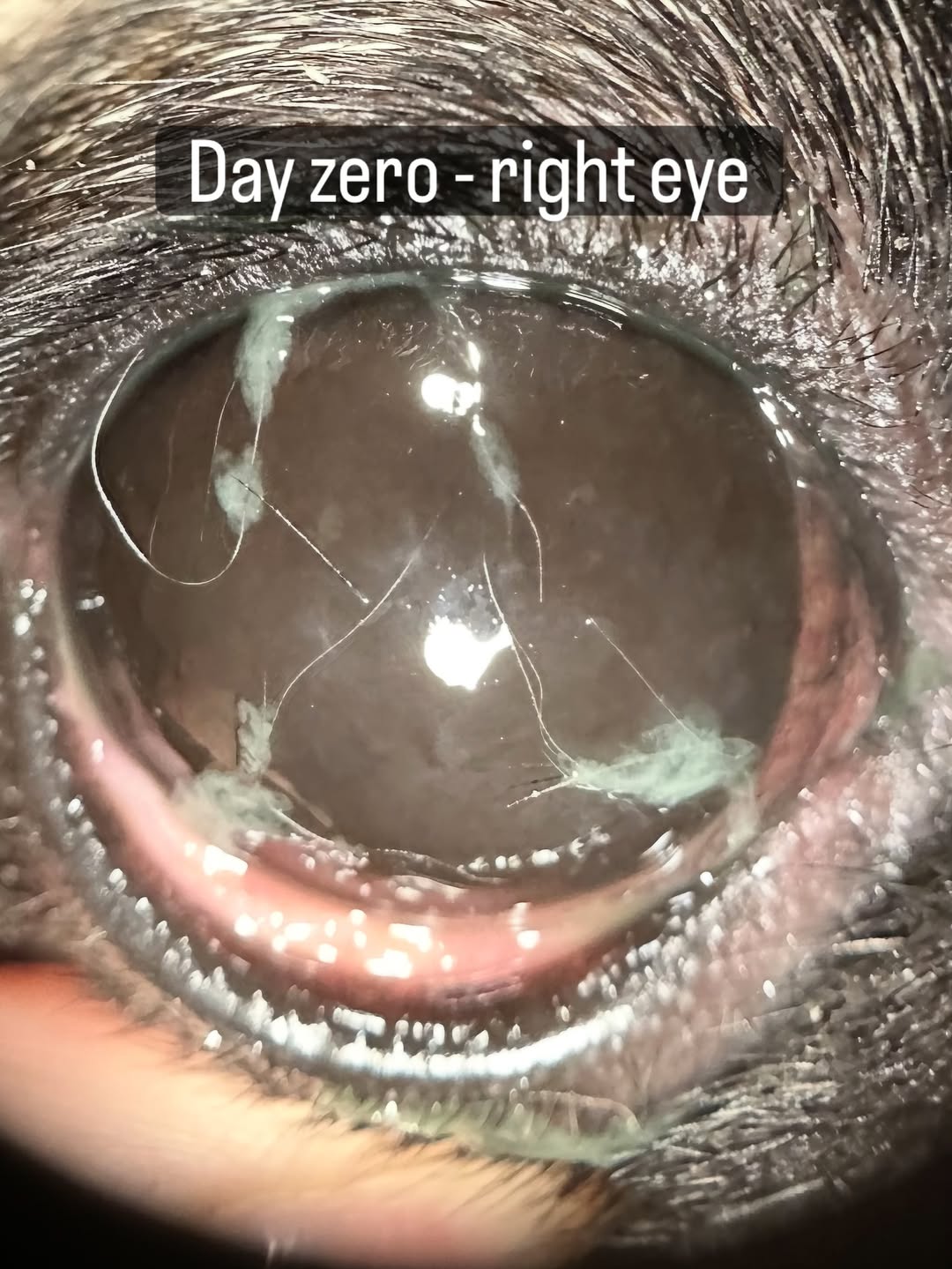
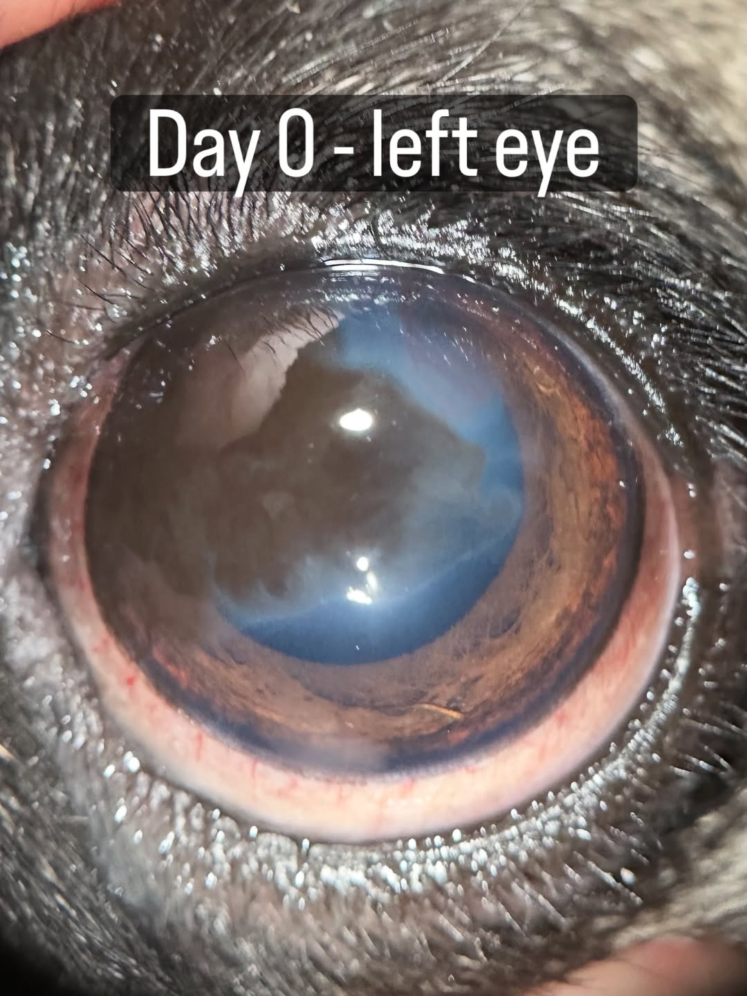
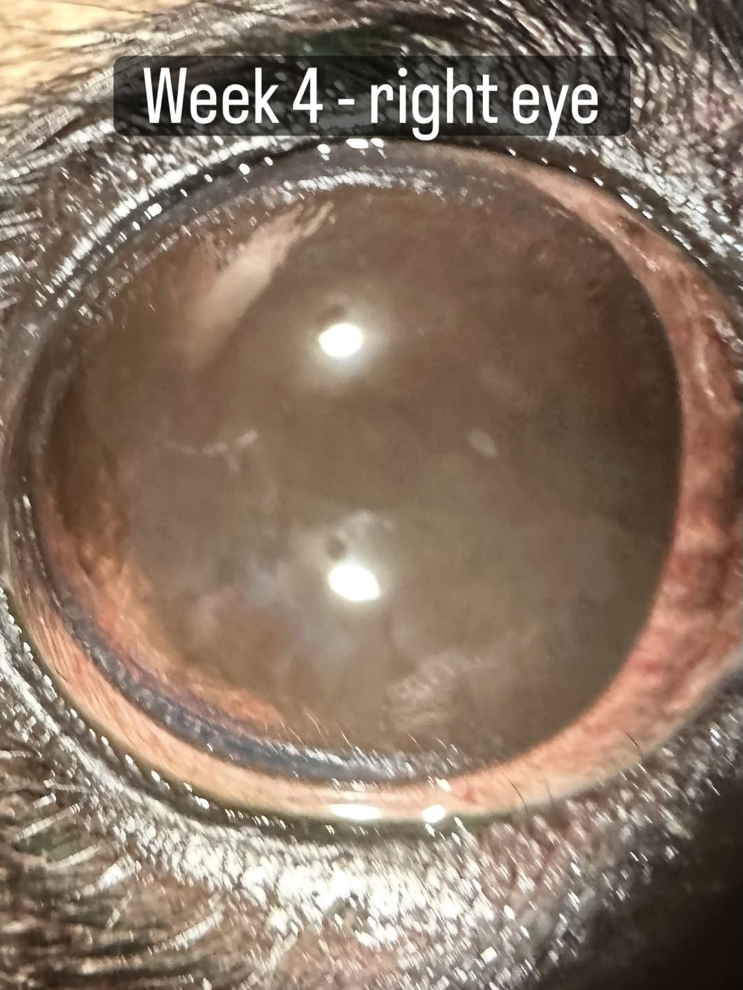
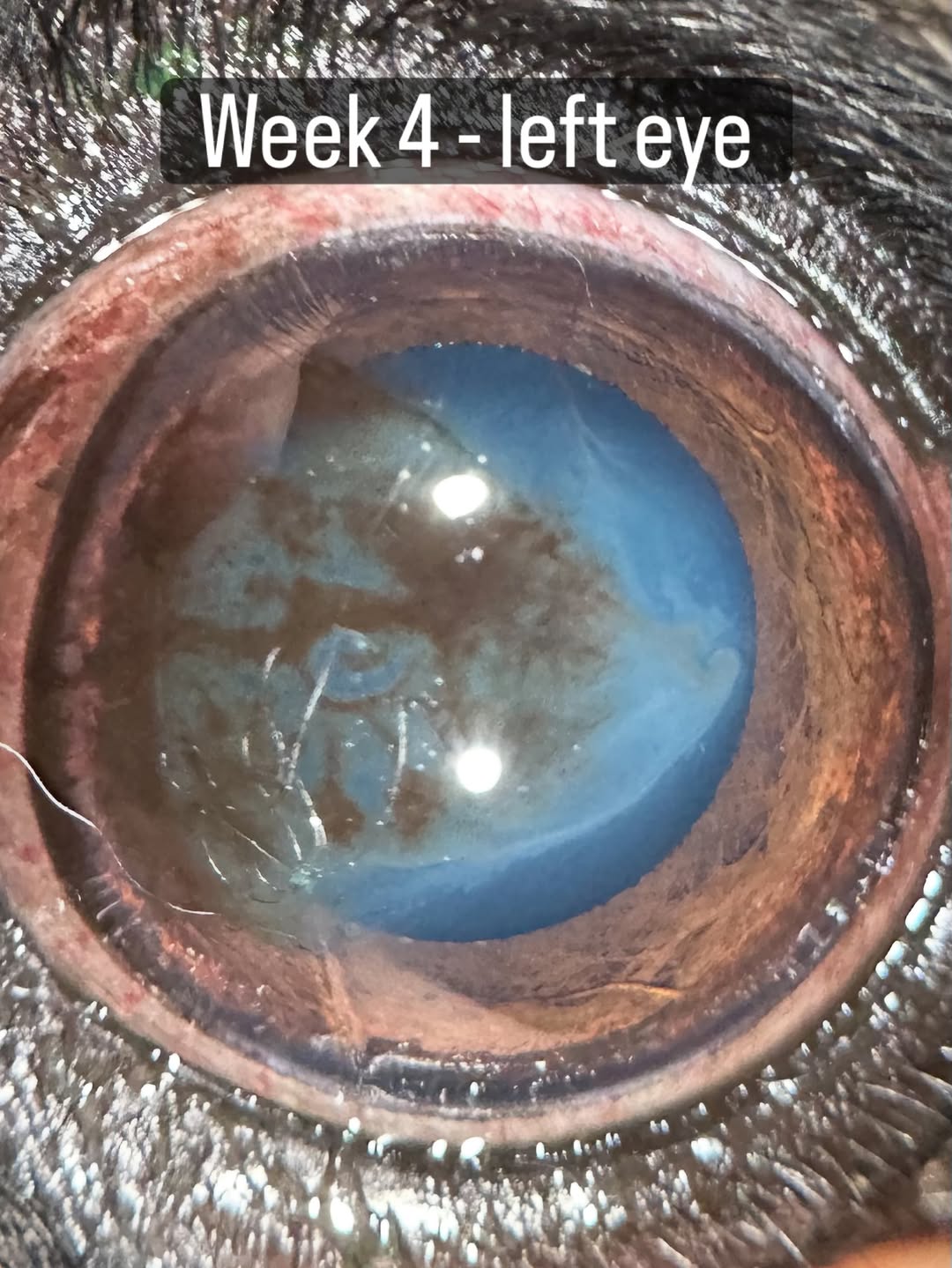
This young Pug was referred for cloudy eyes and lots of goopy eye discharge. As you can see, she has pretty significant corneal pigmentation, with the left eye developing a large wedge of pigment and the right corneal basically completely opaque. She also has very severe dry eye (low tear production) which was a contributing factor. Pigmentary keratitis occurs in brachycephalic dogs commonly due to their wide eyelid fissure and highly exposed eyes, with other conformational factors playing a role.
We have several treatment options ranging from conservative medical therapies with tear stimulants and anti-inflammatory drops (what this owner elected) to surgery to reduce the eyelid fissure and cryotherapy for the corneal pigmentation. While this dog would likely benefit from surgery, they are hoping to avoid anesthesia. We started medication and she has made really good progress in just 1 month as you can see! It’s most apparent in the less severe eye, where the pigment is breaking up and becoming more "wispy". The right eye is also improving, with no more yucky mucoid discharge or redness, and the pigment has started to clear around the edges of the cornea. It's hard to tell in the still images, especially since the patient is SO excited she will barely hold still! I am hopeful we'll continue to get improvement over the next few months. While she will never have a normal cornea in the right eye, if we can prevent worsening and keep her comfortable, her family will be happy!
 |  |
 |  |
We are glad to share a vet case study captured by QuikVue eye imaging adaptor from Dr. Allison Fuchs.
Welcome to the world of brachycephalic breeds and pigmentary keratitis!
This young Pug was referred for cloudy eyes and lots of goopy eye discharge. As you can see, she has pretty significant corneal pigmentation, with the left eye developing a large wedge of pigment and the right corneal basically completely opaque. She also has very severe dry eye (low tear production) which was a contributing factor. Pigmentary keratitis occurs in brachycephalic dogs commonly due to their wide eyelid fissure and highly exposed eyes, with other conformational factors playing a role.
We have several treatment options ranging from conservative medical therapies with tear stimulants and anti-inflammatory drops (what this owner elected) to surgery to reduce the eyelid fissure and cryotherapy for the corneal pigmentation. While this dog would likely benefit from surgery, they are hoping to avoid anesthesia. We started medication and she has made really good progress in just 1 month as you can see! It’s most apparent in the less severe eye, where the pigment is breaking up and becoming more "wispy". The right eye is also improving, with no more yucky mucoid discharge or redness, and the pigment has started to clear around the edges of the cornea. It's hard to tell in the still images, especially since the patient is SO excited she will barely hold still! I am hopeful we'll continue to get improvement over the next few months. While she will never have a normal cornea in the right eye, if we can prevent worsening and keep her comfortable, her family will be happy!
| |
| |