QuikVue Vet Case Share - corneoscleral vessels
We are glad to share a vet case study captured by QuikVue eye imaging adaptor from Dr. Allison Fuchs.
We had a BUSY week friends! Yesterday was a day filled to the brim with equine appointments - fortunately they were all able to stack on the same day and I am so appreciative to be able to see them with a local clinic where we can haul in, have a lovely facility, and stay warm.
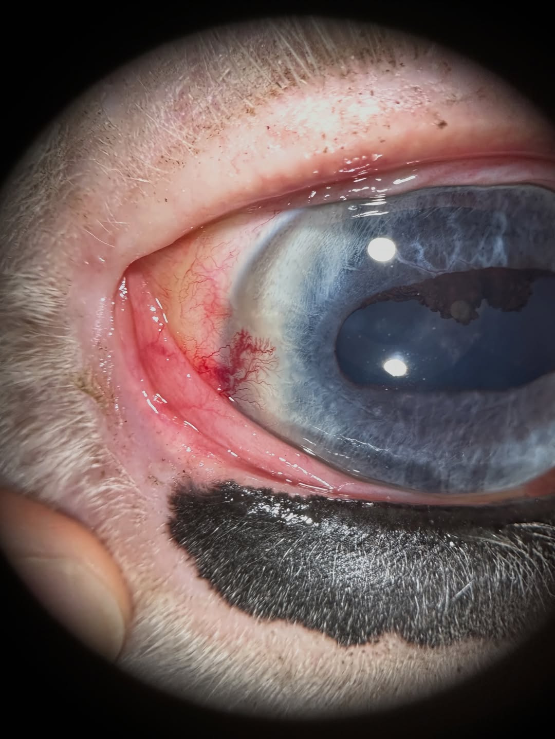
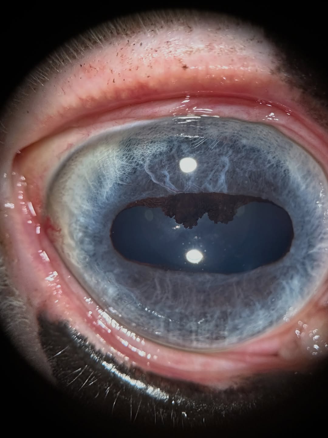
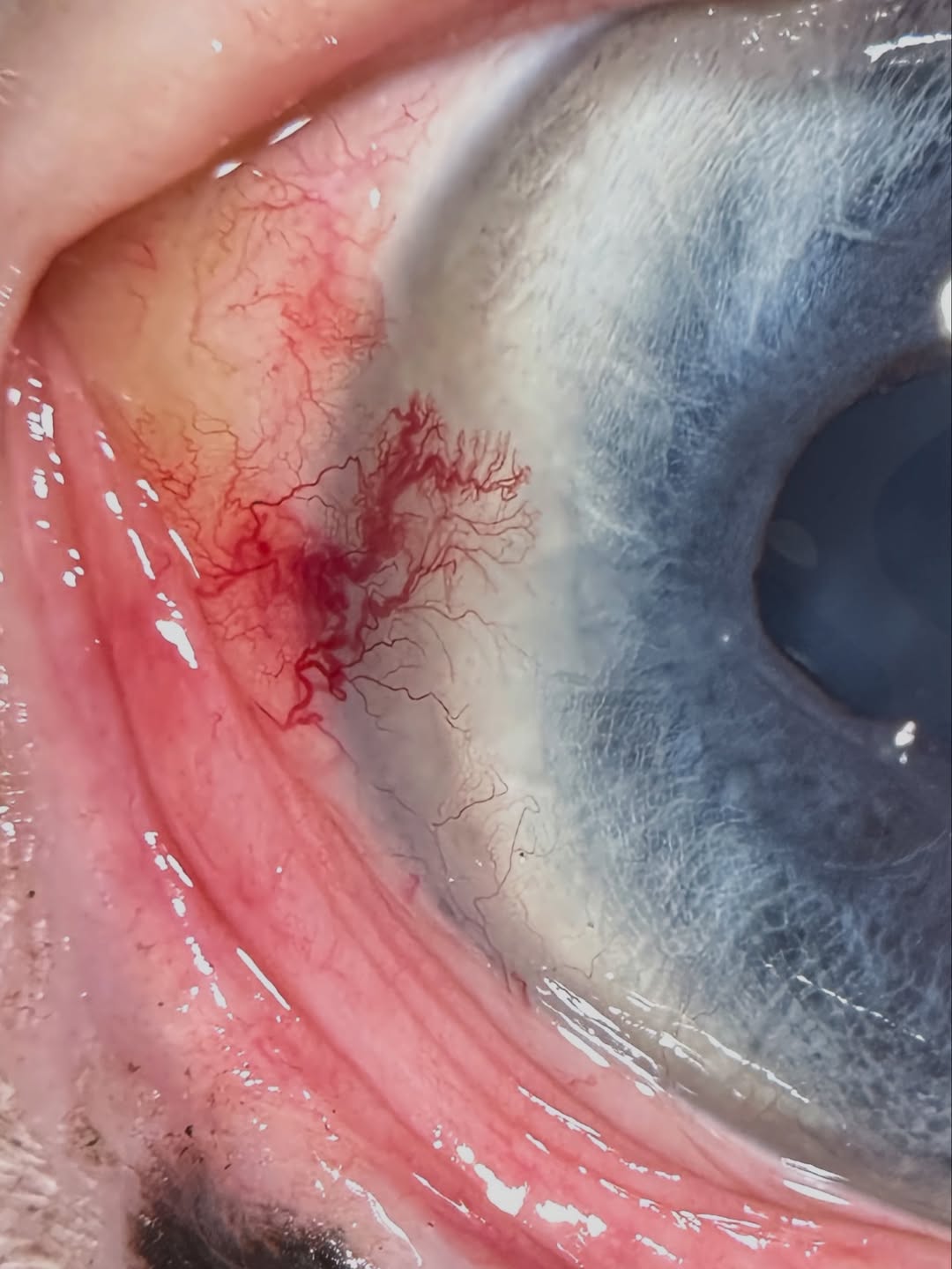
This is one of the horses who is an older guy having some discharge and redness from the right eye. The referring vet noticed the problem and promptly referred. Can you spot it in the first photo? Swipe through for close up images!
This horse has raised, inflamed corneoscleral vessels at the outer corner (lateral limbus) of the eye. This can be a type of immune keratitis, but I am concerned about a cancer called squamous cell carcinoma. This is common in horses around the eyes due to high sun exposure, especially in areas with no pigment. Some breeds are more predisposed due to genetics, like Belgians and Haflingers.
We are planning a surgical keratectomy to remove the affected cornea and sclera, which will also allow us to submit this for biopsy and get a diagnosis. This can often be done with standing sedation and appropriate local anesthesia so we can avoid a general anesthetic episode for what should be a brief procedure. This allows us to minimize risks to the patient and should give an overall better prognosis and outcome!
 |  |  |
We are glad to share a vet case study captured by QuikVue eye imaging adaptor from Dr. Allison Fuchs.
We had a BUSY week friends! Yesterday was a day filled to the brim with equine appointments - fortunately they were all able to stack on the same day and I am so appreciative to be able to see them with a local clinic where we can haul in, have a lovely facility, and stay warm.
This is one of the horses who is an older guy having some discharge and redness from the right eye. The referring vet noticed the problem and promptly referred. Can you spot it in the first photo? Swipe through for close up images!
This horse has raised, inflamed corneoscleral vessels at the outer corner (lateral limbus) of the eye. This can be a type of immune keratitis, but I am concerned about a cancer called squamous cell carcinoma. This is common in horses around the eyes due to high sun exposure, especially in areas with no pigment. Some breeds are more predisposed due to genetics, like Belgians and Haflingers.
We are planning a surgical keratectomy to remove the affected cornea and sclera, which will also allow us to submit this for biopsy and get a diagnosis. This can often be done with standing sedation and appropriate local anesthesia so we can avoid a general anesthetic episode for what should be a brief procedure. This allows us to minimize risks to the patient and should give an overall better prognosis and outcome!
| | |