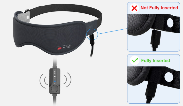
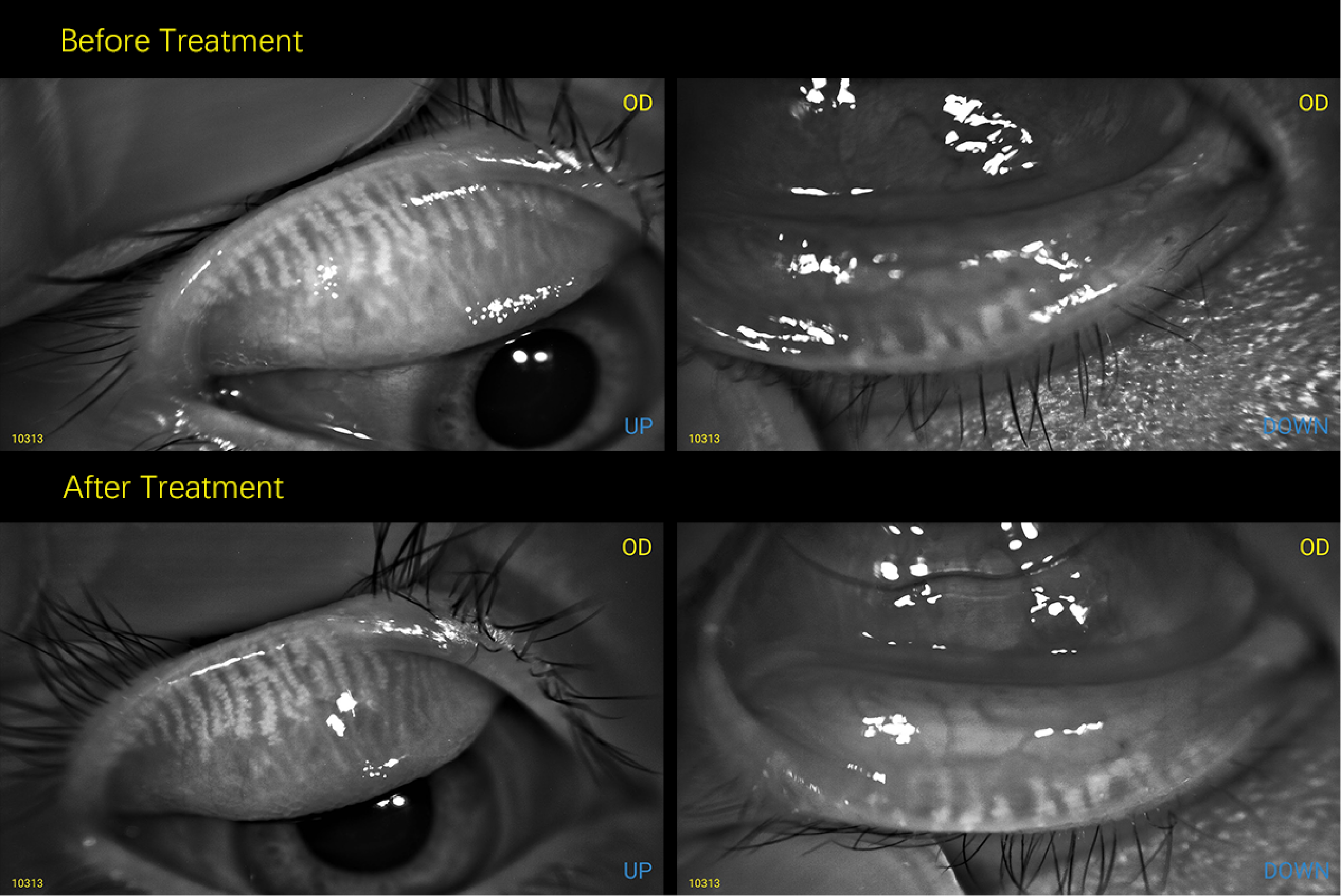
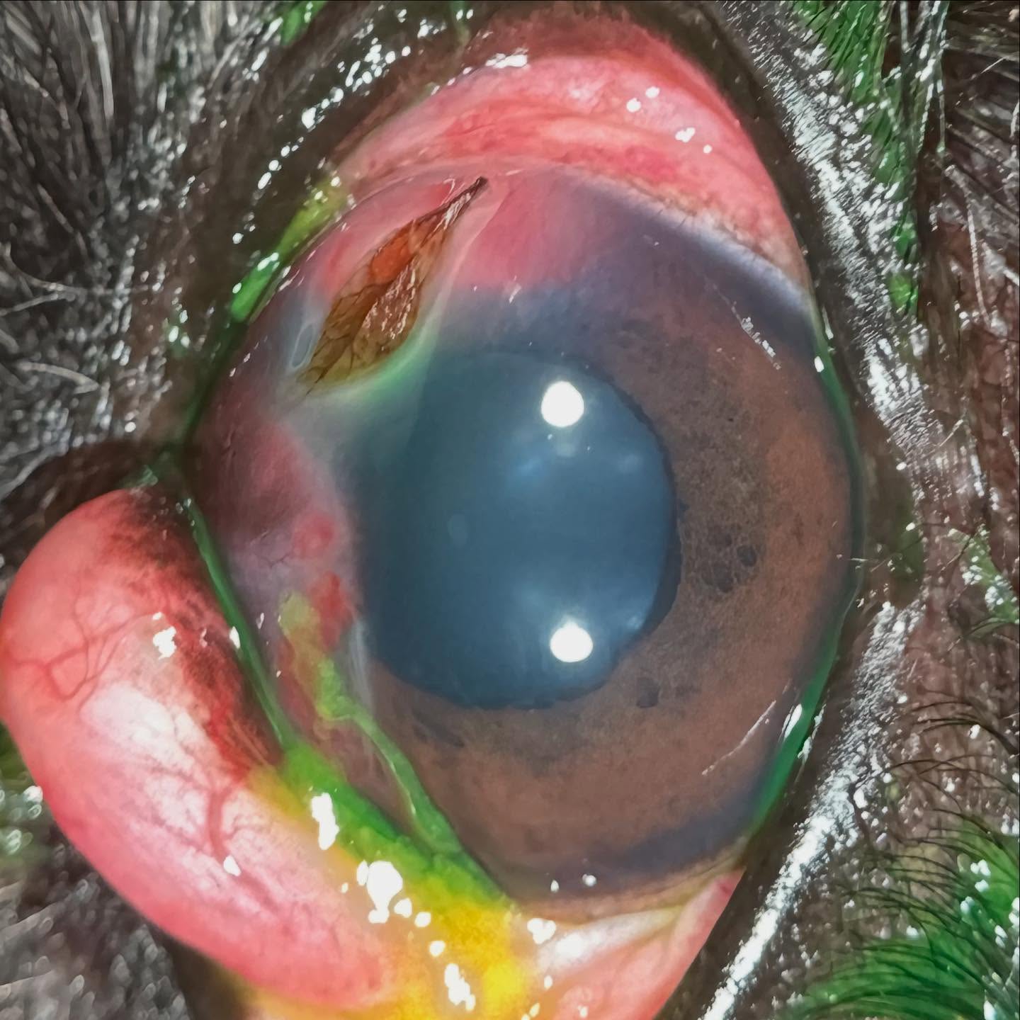
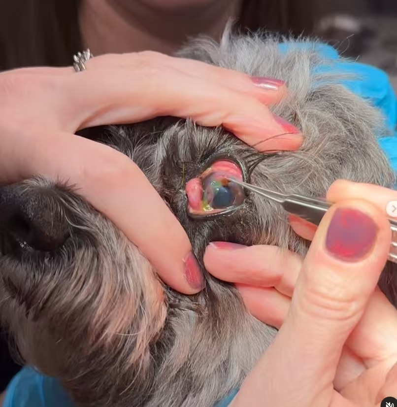
QuikVue Vet Case Study—Corneal Foreign Body in a Pup
We are glad to share a vet case study captured by QuikVue eye imaging adaptor from Dr.Allison Fuchs.
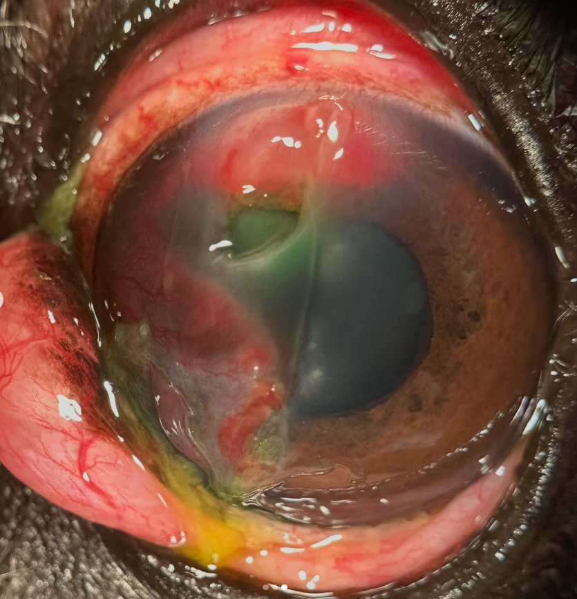
Who would be-leaf it?? See what I did there? This sweet pup was referred to see me emergently today due to concern for a corneal laceration. He had been experiencing ocular discomfort for about 2 weeks, which was much worse the last day or two. A dark spot was noted in the cornea of the left eye, in a large divot. He also has chronic prolapse of the third eyelid gland (also known as cherry eye), and corneal inflammation (keratitis). His ophthalmic exam revealed a LEAF embedding its way into the cornea! We removed the leaf with topical anesthesia, and are going to treat the resulting deep corneal ulcer medically. Fingers crossed that this pup heals up and then we can focus on his third eyelids and chronic corneal disease.
 |  |  |
We are glad to share a vet case study captured by QuikVue eye imaging adaptor from Dr.Allison Fuchs.
Who would be-leaf it?? See what I did there? This sweet pup was referred to see me emergently today due to concern for a corneal laceration. He had been experiencing ocular discomfort for about 2 weeks, which was much worse the last day or two. A dark spot was noted in the cornea of the left eye, in a large divot. He also has chronic prolapse of the third eyelid gland (also known as cherry eye), and corneal inflammation (keratitis). His ophthalmic exam revealed a LEAF embedding its way into the cornea! We removed the leaf with topical anesthesia, and are going to treat the resulting deep corneal ulcer medically. Fingers crossed that this pup heals up and then we can focus on his third eyelids and chronic corneal disease.
| | |