QuikVue Vet Case Study
We are glad to share a vet case study captured by QuikVue eye imaging adaptor from Dr.Allison Fuchs.
This sweet little Boston presented emergently last week for squinting and a corneal ulcer. As you can see in the first image, he has an ulcer with some irregular material in it that turned out to be a corneal foreign body with plant material! Once removed, there is an infected ulcer that is getting quite deep. We started aggressive medical therapy with topical antibiotics and serum, as well as oral antibiotics and pain medication. After just 5 days the ulcer is almost completely healed to a scar and the infection is almost cleared. This is great progress and a big relief for this dog and his family!
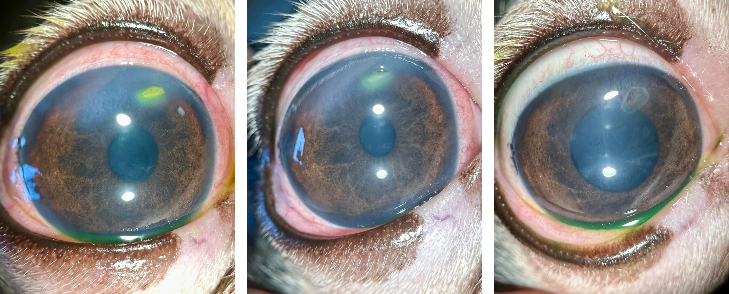 |
We are glad to share a vet case study captured by QuikVue eye imaging adaptor from Dr.Allison Fuchs.
This sweet little Boston presented emergently last week for squinting and a corneal ulcer. As you can see in the first image, he has an ulcer with some irregular material in it that turned out to be a corneal foreign body with plant material! Once removed, there is an infected ulcer that is getting quite deep. We started aggressive medical therapy with topical antibiotics and serum, as well as oral antibiotics and pain medication. After just 5 days the ulcer is almost completely healed to a scar and the infection is almost cleared. This is great progress and a big relief for this dog and his family!
|