QuikVue Vet Case Study—Progressive Rod-Cone Degeneration, PRCD
We are glad to share a vet case study captured by QuikVue eye imaging adaptor from Dr.Allison Fuchs.
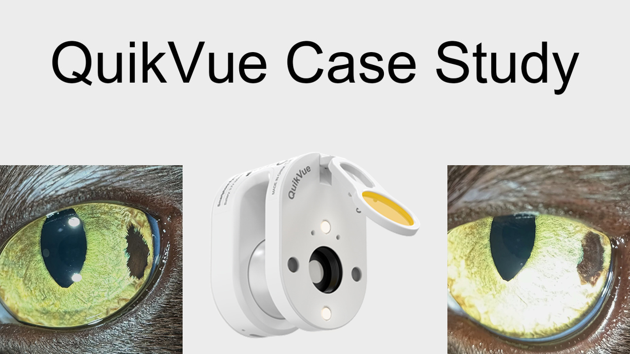
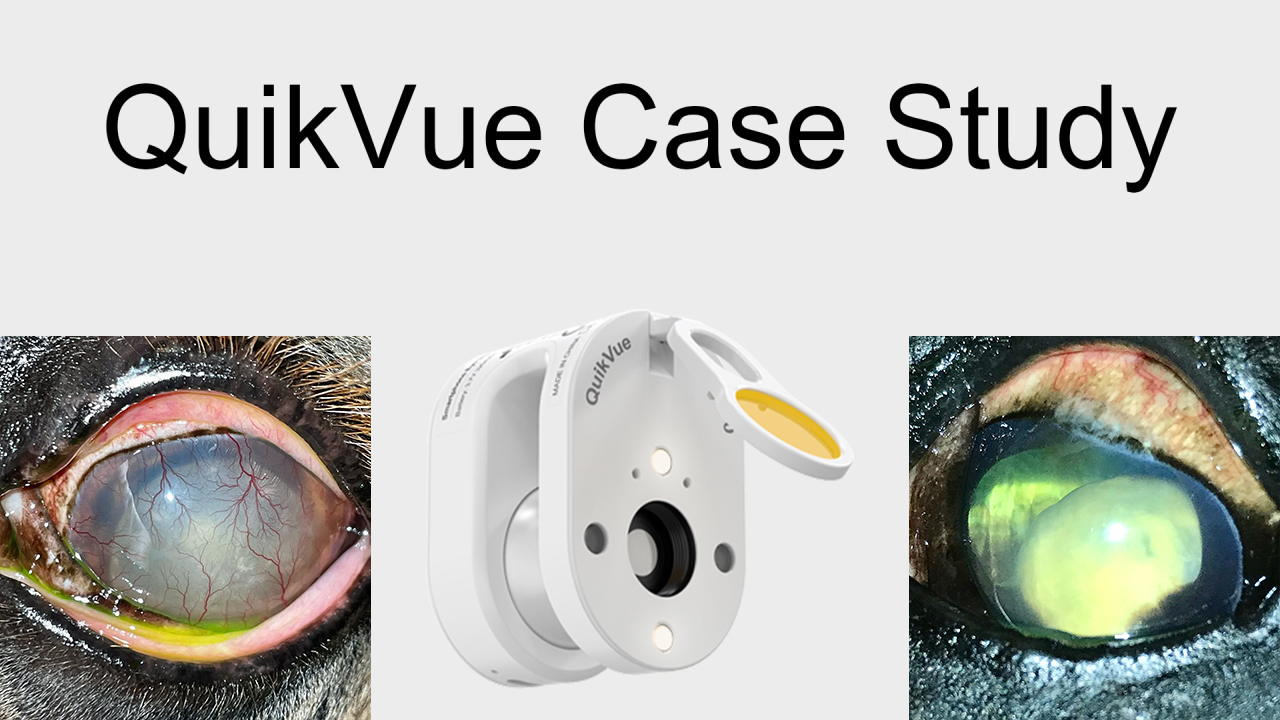
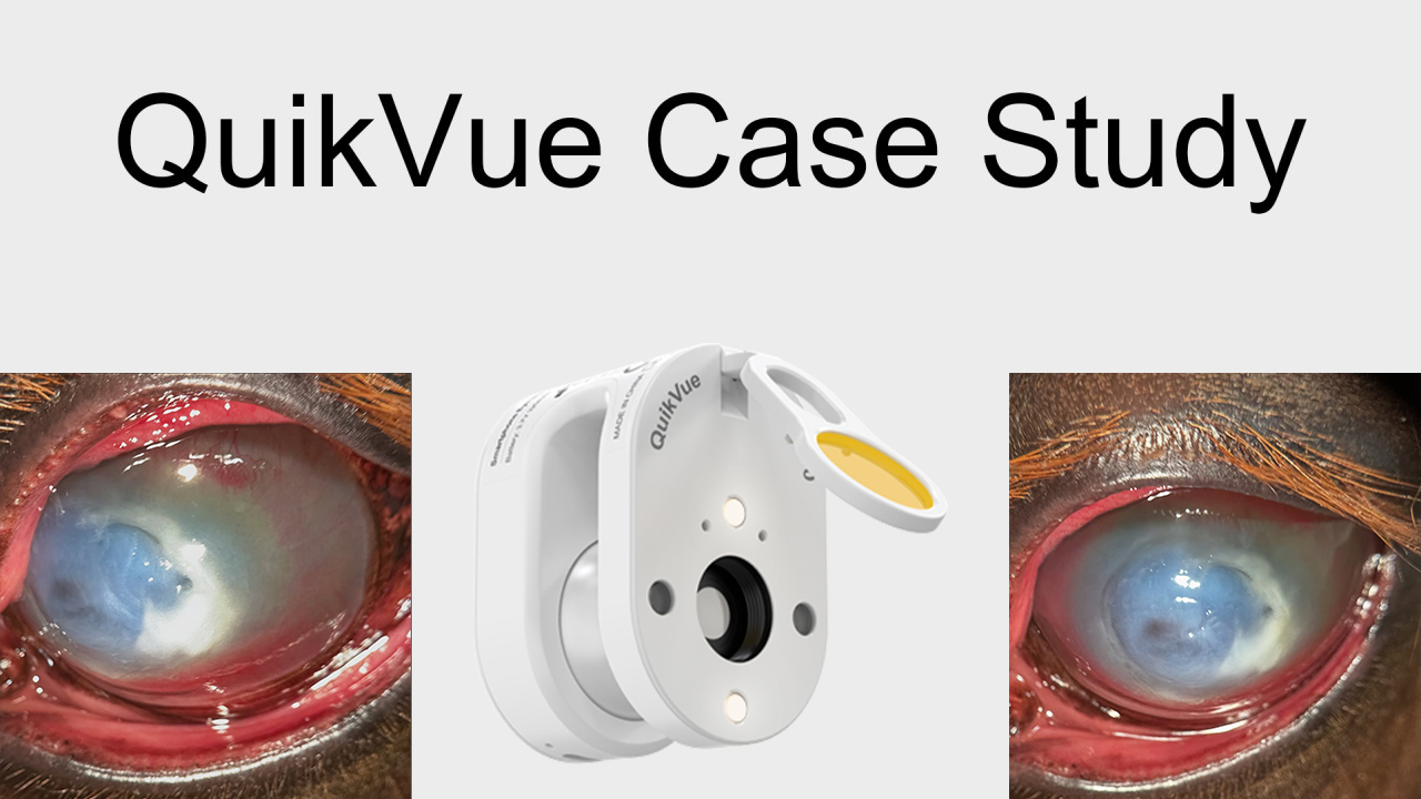
This sweet pittie presented to us for possible vision loss last week. The owner hasn’t really noticed much change at home, but when she was boarded she had trouble getting around, especially at nighttime. We did administer dilating eye drops during this appointment. She has some vision, but her responses are reduced in normal room light and absent in dim light. This dog does have early cataracts, which are primarily affecting the posterior cortex of the lens. Fundic exam was easily performed, which means if everything is working properly the dog should be able to see out. The history, reduced menace response, and progressive cortical cataracts should be a red flag for retinal degeneration. On fundic exam, this dog’s retinas were at an advanced stage of atrophy. You can see the optic nerve is pale, the retinal blood vessels are thin (attenuated), and the tapetum is EXTRA shiny (hyperreflective), which indicates thinning of the neurosensory retina. All is consistent with progressive rod-cone degeneration, aka PRCD or PRA. This is a group of inherited retinal degenerations that can affect just about any breed of dog, and rarely cats. Cataract development is common due to release of toxic factors from the retina. While cataract surgery can be helpful in some cases, it is usually a short term fix as the retinal degeneration is not treatable.
 |  |