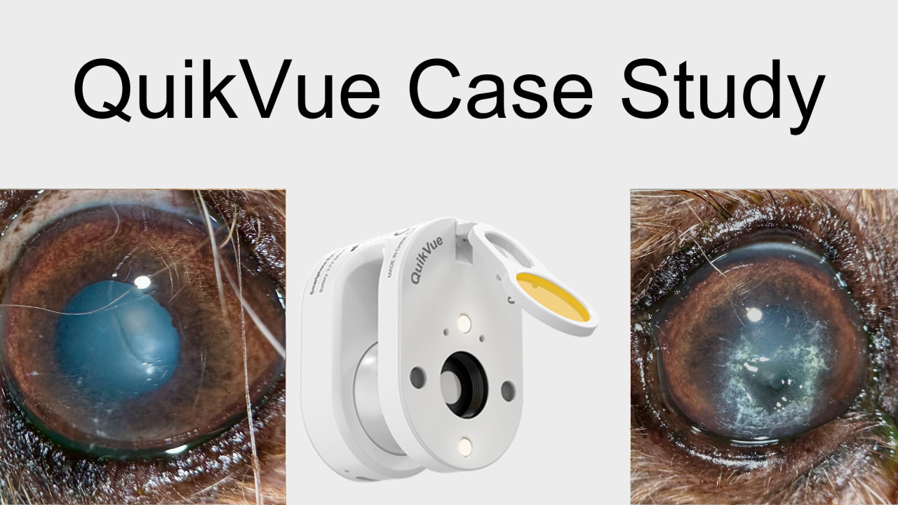
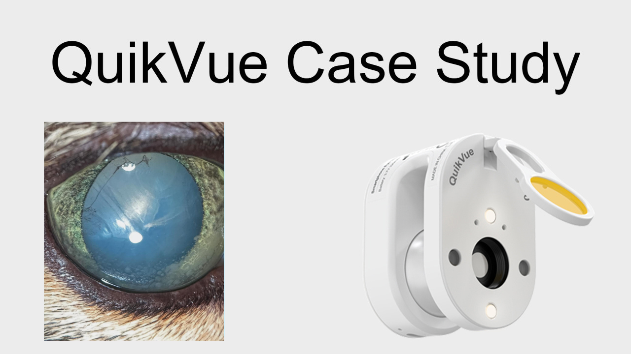
QuikVue Vet Case Study—Feline Iris Melanosis
We are glad to share a vet case study captured by QuikVue eye imaging adaptor from Dr.Allison Fuchs.
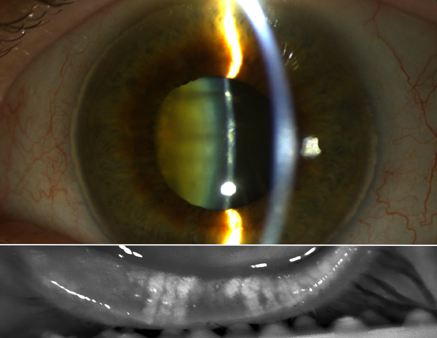
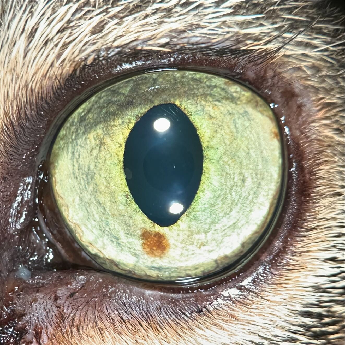
This is an 11yr old cat with progressive iris pigmentation. Her intraocular pressure is normal and there is no active inflammation inside the eye. She is visual and comfortable. The pigment spot has been present for at least 6 months and the owners have followed with photos at home and noted growth. What is this and what do we do about it? I find this condition challenging because we have so little solid information about how it behaves! This is FDIM, or feline diffuse iris melanosis/melanoma. There is no real way to tell without histopathology whether the change is benign or malignant. There are reports of metastatic disease from iris melanoma in cats, but the reported rates range from 16-63% which is wildly unhelpful. Some people advocate for early enucleation. Personally I offer a range of options depending on the cat’s age, rate of progression, the appearance of the lesions, and the owner’s motivation. We can monitor (watch and wait), take an iris biopsy, perform trans-corneal laser ablation to try to slow down the growth, or remove the eye. I also offer staging (chest x-rays and abdominal ultrasound) prior to surgery, especially in more advanced cases. This is a thankfully very early case with relatively slow progression, and may be entirely benign! We are going to monitor her for now, plan to recheck with me in about 4 months, and will probably proceed to laser if it is changing or the owners want to be more aggressive.
 |