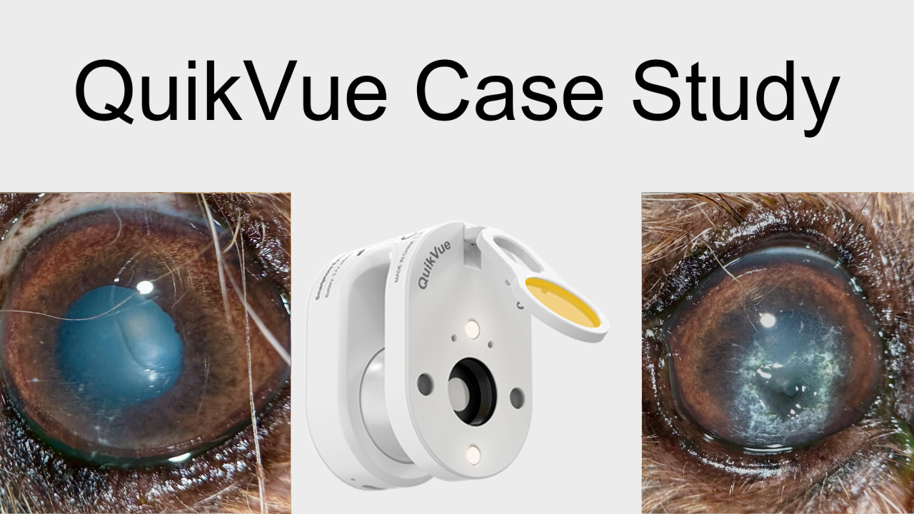
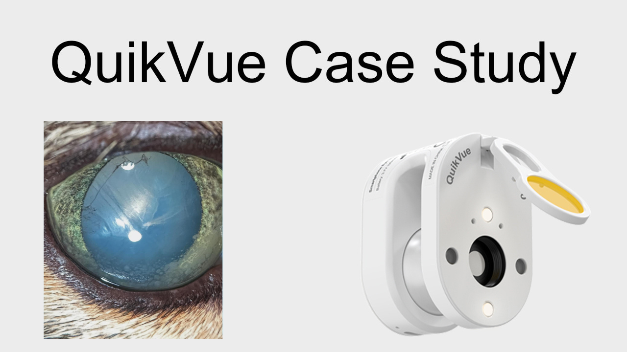
QuikVue Vet Case Study—Corneal Ulcer with Intraocular Hemorrhage
We are glad to share a vet case study captured by QuikVue eye imaging adaptor from Dr.Allison Fuchs.
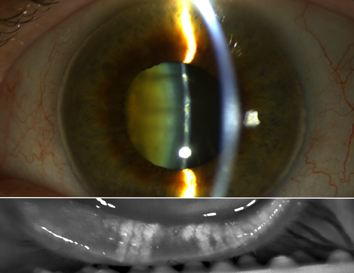
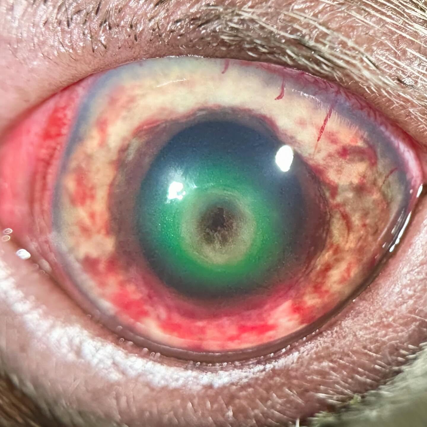
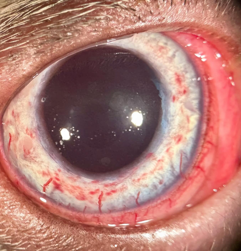
Can you guess what concerned me most about this patient? I’ll give you a hint, it’s not the cornea! This young dog was referred for a deep ulcer, and he does indeed have a gnarly descemetocele. Any other day we’d be heading to surgery to graft this, either with a conjunctival pedicle or corneal-conjunctival transposition. However, this dog is bleeding into his iris. In BOTH eyes. This puts a hard stop on surgery, at least for me, until we figure out why and what his risk is of spontaneous bleeding. He is also a brachycephalic breed and already at higher risk of peri-anesthetic complications! We did a thorough systemic workup, including coagulation tests and platelet function (yes I had to do a BMBT!) and his tests all came back relatively normal. Good news, but also bad because it means we don’t have a cause. Due to the risks and in absence of finding an underlying etiology for the bleeding, we elected to start aggressive medical therapy for the eye, and systemic therapy with an antibiotic that covers most tick-borne diseases. Thankfully he is improving rapidly, both in regards to the ulcer and the bleeding. Super weird case, and once again a reminder of evaluating the entire patient even when faced with a scary looking eyeball!
 |  |