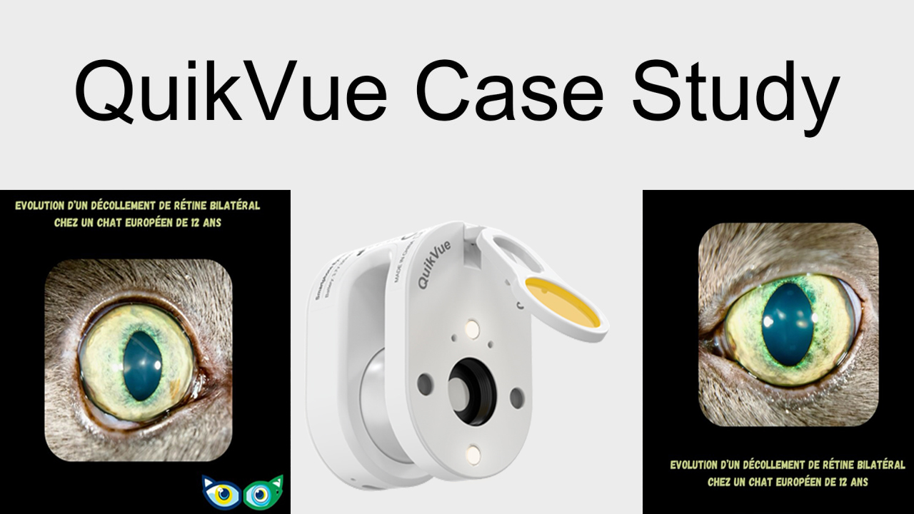
QuikVue Vet Case Share-Bilateral Amaurosis in an European Cat
We are glad to share a vet case study captured by QuikVue eye imaging adaptor from Dr Vet Viroux.
A male neutered 8-year-old European cat was presented for bilateral mydriasis without loss of vision.
Clinical examination: No abnormalities were found.
Ophthalmological examination: The menace response and Dazzle reflex were both positive, but the pupillary light reflexes were negative. The iris showed no atrophy, and the fundus was normal.
Neurological involvement was suspected, and an MRI exam was recommended but not performed at that time.
Three months later, the cat was presented again with disorientation and vision loss progressing over several days.
Clinical examination: No abnormalities were found, and blood pressure was normal at 150 mmHg.
Ophthalmological examination: This time, a negative menace response was observed on the right and diminished on the left, with a negative Dazzle reflex. The rest of the examination was unremarkable.
MRI exam was recommended again to explore intracranial causes, which was accepted this time. The MRI revealed a large mass invading the pituitary fossa and optic foramina, along with subtle signs of intracranial hypertension.
Explanation: The nerve fibers responsible for the pupillary light reflex decussate after the optic nerve decussation, caudal to the optic chiasm. This anatomical feature could explain why bilateral mydriasis occurred before vision loss, as these neural pathways do not participate in vision.